Pulmonary Sequestration

  • Etiology: nonfunctional dysplastic pulmonary parenchyma lung tissue not connected to bronchial tree or pulmonary artery, comes from accessory lung bud, if it develops early before pleura develops is intralobar, if it develops later after pleura develops is extralobar
  • Imaging: most commonly in lower lobes (left > right), arterial blood supply from subdiaphragmatic aorta / celiac trunk / splenic artery, only contain air when infected, 15% are ectopic – suprarenal / mediastinum / pericardial / neck
    — Intralobar (75%) – no separate pleural investment (within visceral pleura), pulmonary venous drainage to left atrium (normal drainage)
    — Extralobar (25%) – separate pleural investment (outside visceral pleura), systemic venous drainage to vena cava or azygous vein, 15% ectopic – suprarenal / mediastinal / pericardial / neck
  • Clinical: intralobar present in older child with recurrent lower lobe pneumonias, 60% of extralobar sequestrations are associated with congenital abnormalities (congenital diaphragmatic hernia, cardiac anomalies, congenital pulmonary airway malformation = hybrid lesion)

Radiology Cases of Pulmonary Sequestration

Radiology Cases of Intralobar Pulmonary Sequestration

Radiology Cases of Extralobar Pulmonary Sequestration

CXR and MRI of pulmonary sequestration
CXR shows a large round opacity in the left lower lobe that abuts the diaphragm. Coronal T1 MRI with contrast of the chest shows a uniformly enhancing mediastinal mass that extends into the retrocrural regions of the chest bilaterally and that has a feeding vessel arising from the aorta.
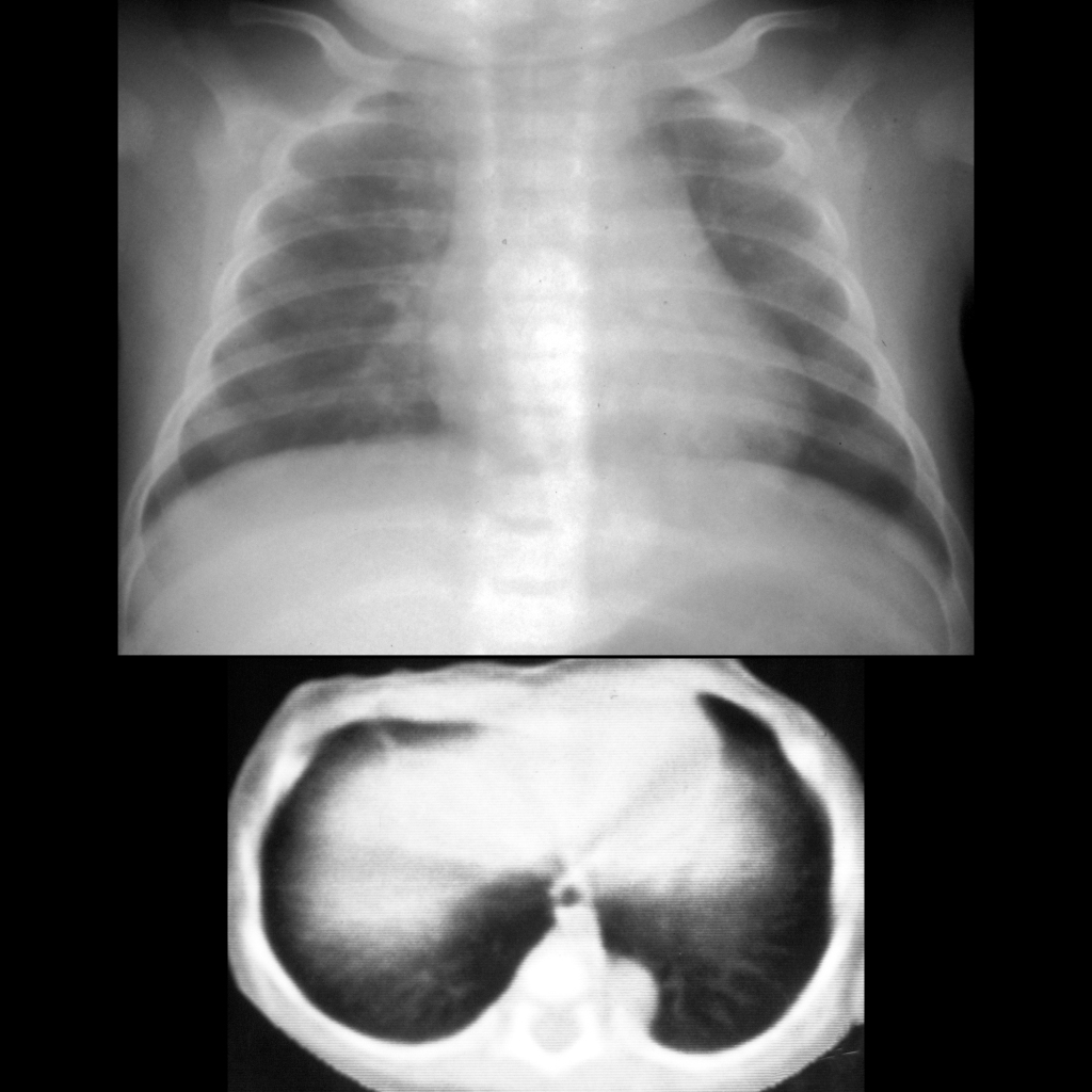
CXR and CT of extrapulmonary sequestration
CXR AP (above) shows a round density projecting to the left of the 8th and 9th thoracic vertebral bodies. Axial CT without contrast of the chest (below) better shows a solid left paraspinal mass. In the operating room it was seen to lay outside the lung and to have its arterial supply coming from the aorta.

Gross Pathology Cases of Pulmonary Sequestration

Gross pathological image of extralobar pulmonary sequestration
Gross pathological images show a right-sided subdiaphragmatic / suprarenal mass similar in appearance to the lung above it (above left). When the aorta was opened (above right), the orifice of an arterial vessel supplying the mass was seen coming off of the aorta just above the tip of the upper hemostat on the right side of the aorta. The relationship of the feeding vessel off of the aorta to the mass is more clearly demonstrated on the specimen image (below).
Pathology image of pulmonary sequestration
Gross pathological image shows a left-sided supradiaphragmatic mass separate from the left lung. There was a feeding vessel connecting it to the aorta.