- Etiology: occurs over 24-36 hours: hyperplasia of lymphoid follicles / parasites / neuroendocrine (carcinoid) tumor -> distension of appendix -> ischemic mucosal damage -> bacterial overgrown and wall invasion -> transmural inflammation -> perforation
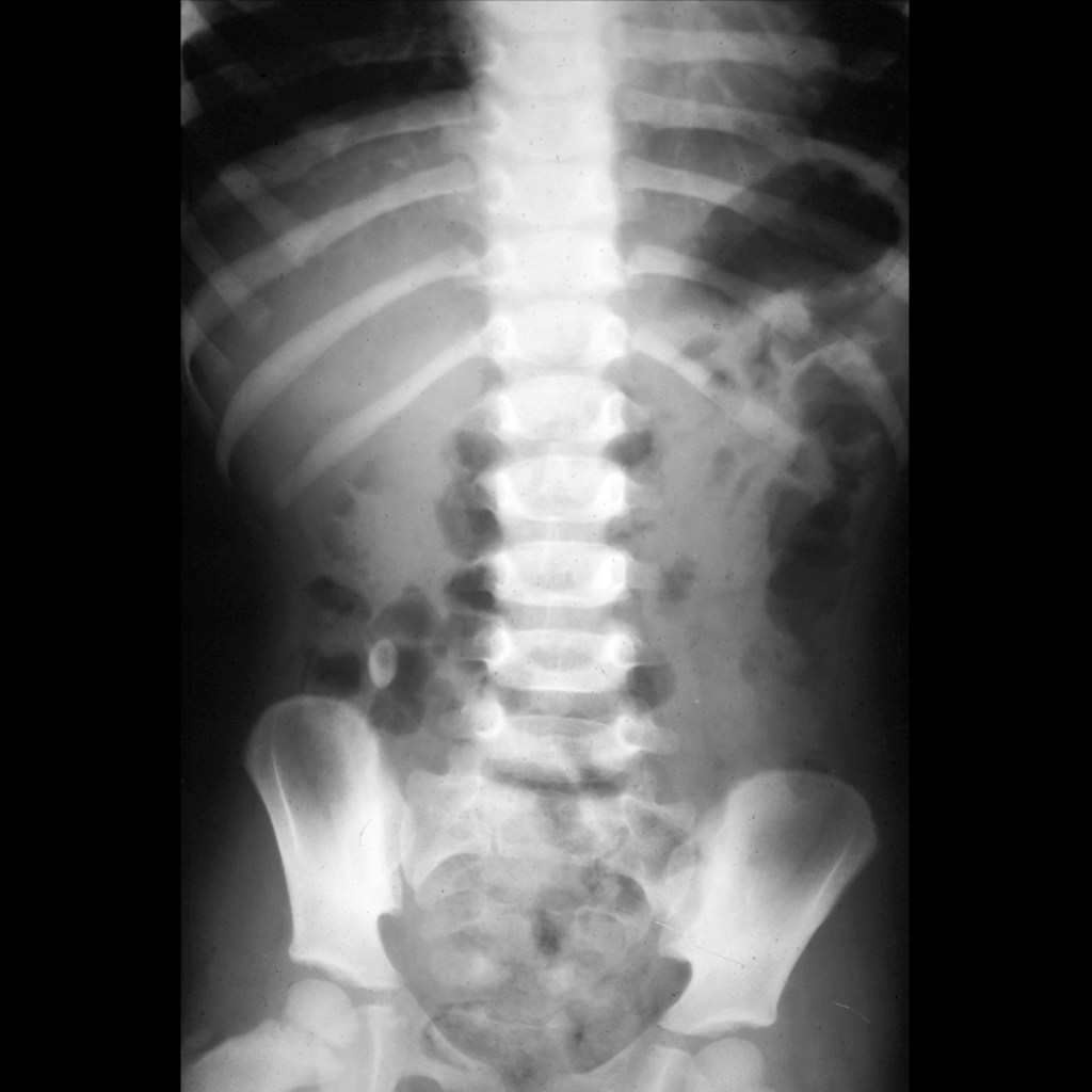
- AXR: appendicolith in 15%, may have distal small bowel obstruction
- US Normal appendix: compressible blind ending tubular structure originating from base of cecum that does not have peristalsis, transverse diameter is < 6 mm, no wall thickening, central echogenic line which is acoustic reflection from collapsed luminal interface, 80% draped over iliac vessels
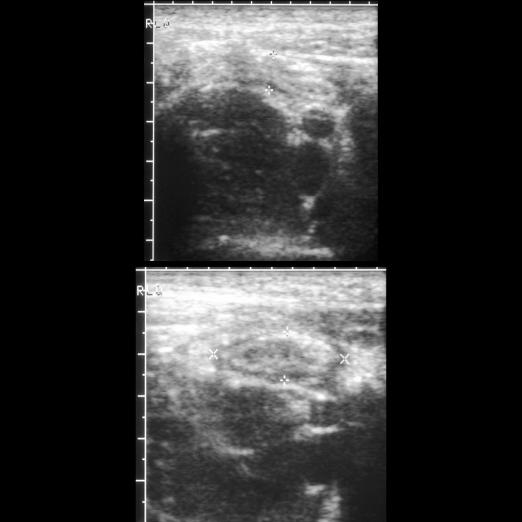
— Note: terminal ileum can mimic appendix but has hypoechoic folds and has peristalsis - US Acute appendicitis: noncompressible blind ending tubular structure with transverse diameter > 6 mm, appendiceal wall thickness > 3 mm, hyperemia on color doppler US, periappediceal hypoechoic halo from wall edema, periappendiceal hyperechogenicty of periappendiceal fat in mesentery from periappendiceal edema, appendicolith
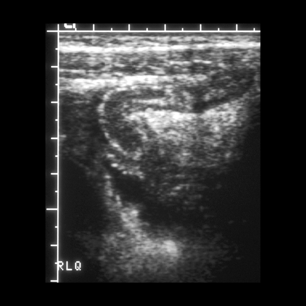
— Note: diameter should not be only criteria, some say appendiceal transverse diameter of 6-8 mm is indeterminate for acute appendicitis and that appendiceal transverse diameter of > 8 mm is diagnostic of acute appendicitis - US Perforated appendicitis: appendix may not be seen or may be decompressed with diameter < 6 mm, phlegmon with poorly defined bowel loops in right lower quadrant with increased echogenicity, mass of mixed echogenicity, focal bowel wall thickening, intraperitoneal fluid, loculated fluid, frank abscess
- CT Acute appendicitis: diameter > 8 mm (diameter 6-8 mm is indeterminate), enhancing, appendicolith in 30%, periappendiceal fat stranding, periappendiceal fluid / abscess are signs of perforation
- MR Normal appendix: less than 7 mm, no periappendiceal changes, paucity of fat can make visualization difficult so lack of inflammatory changes implies a normal exam
- MR Acute appendicitis: diameter > 7 mm, periappendiceal inflammation, focus of diminished signal intensity represents and appendicolith
- Clinical:
— Simple appendicitis: periumbilical pain that localizes to right lower quadrant with fever and vomiting is seen in < 50% of patients, 33% of patients have nonspecific symptoms
— Perforated appendicitis: pain relief then more generalized pain and fever and generalized peritonitis
Radiology Cases of Appendicitis
Radiology Cases of Normal Appendix

Radiology Cases of Acute Appendicitis With Appendicolith


Radiology Cases of Acute Appendicitis


Radiology Cases of Acute Appendicitis in Morgagni Hernia

Radiology Cases of Acute Appendicitis and Carcinoid Tumor of the Appendix

Radiology Cases of Perforated Appendicitis


Radiology Cases of Perforated Appendicitis With Appendicolith

Surgery Cases of Appendicitis

Gross Pathology Cases of Appendicitis

Histopathology Cases of Appendicitis


