- Etiology:
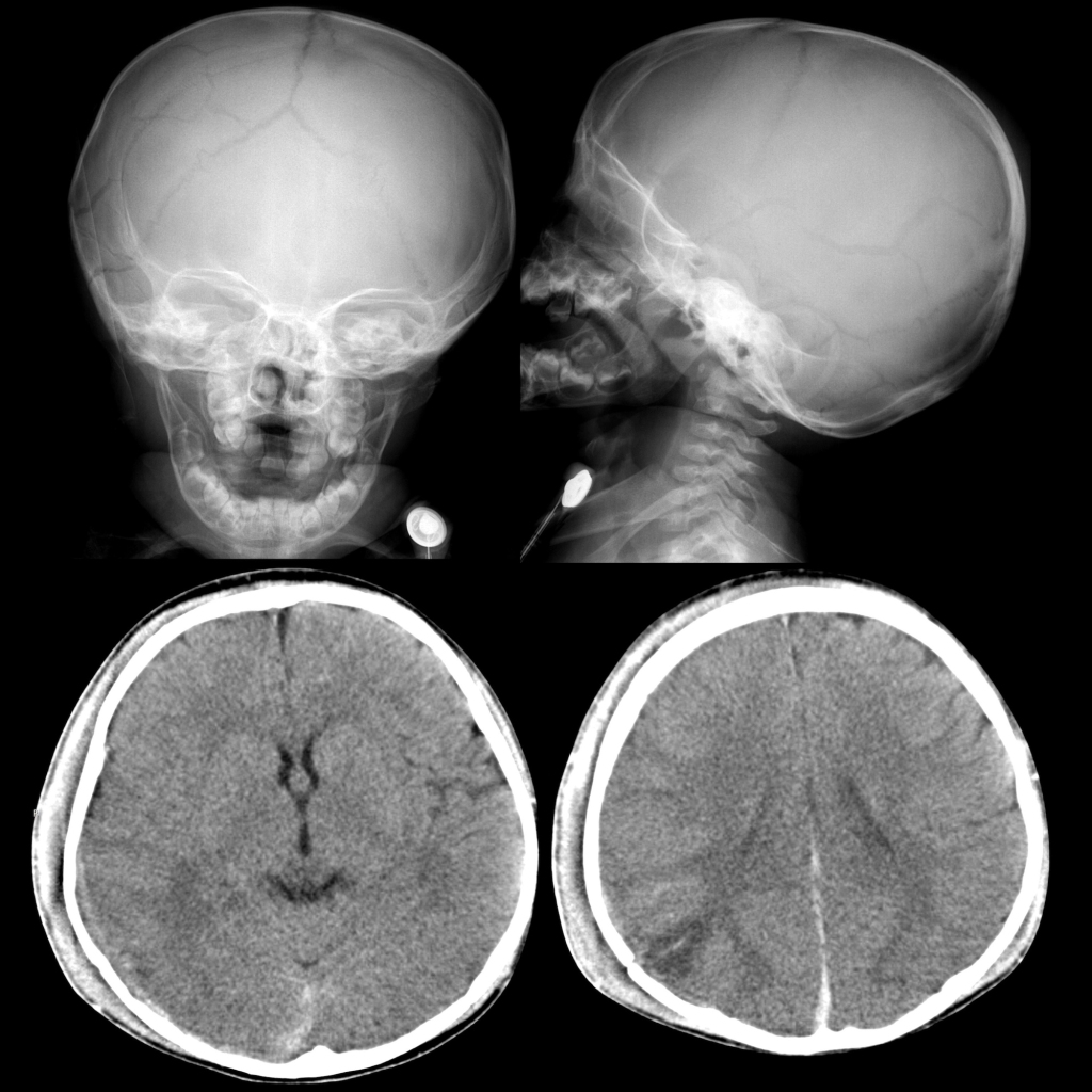
— Direct trauma -> skull fracture (can be multiple, complex), contusion (coup/contrecoup)
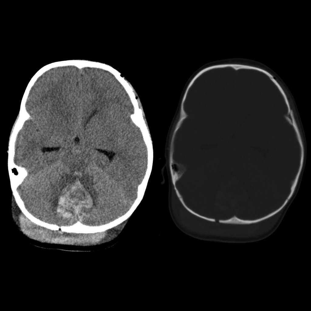
— Shaking -> rotational injury, subdural hematoma (interhemispheric), subarachnoid hemorrhage, contusions, white matter tear/laceration, cervicomedullary stretch -> apnea – > hypoxia / ischemia - Imaging:
— Subdural hematoma is most common intracranial manifestation
— Subdural hematoma can resolve rapidly due to washout from cerebrospinal fluid and redistribution
— Chronic subdural hematoma with enhancing membrane can be seen at around 1 week
— Spontaneous rebleeding into chronic subdural hematoma is rare in infants unless there is underlying cerebral atrophy or ventriculoperitoneal shunt with low intracranial pressure
— Mixed density subdural hematoma is due to acute cerebrospinal fluid from tear in arachnoid membrane mixing with acute blood, early clot retraction, acute unclotted blood, acute hemorrhage into chronic subdural hematoma - Note: may have associated hemorrhage in the spinal canal
- DDX: benign enlargement of the subarachnoid space can mimic chronic bilateral subdural hematomas
- Complications: intracranial herniation
- Clinical: intracranial injury is the most common cause of death
Radiology Cases of Child Abuse Neurological Injury
Radiology Cases of Child Abuse Skull Fracture

Radiology Cases of Child Abuse Subdural Hematoma


Radiology Cases of Child Abuse Diffuse Cerebral Edema


