A Pediatric Radiology Textbook and Pediatric Radiology Digital Library
Pediatric Hangman Fracture
Etiology: trauma
Imaging: fractures through bilateral C2 pedicles results in avulsion of C2 body from arch with anterior translation of C2 on C3
Radiology Cases of Hangman Fracture
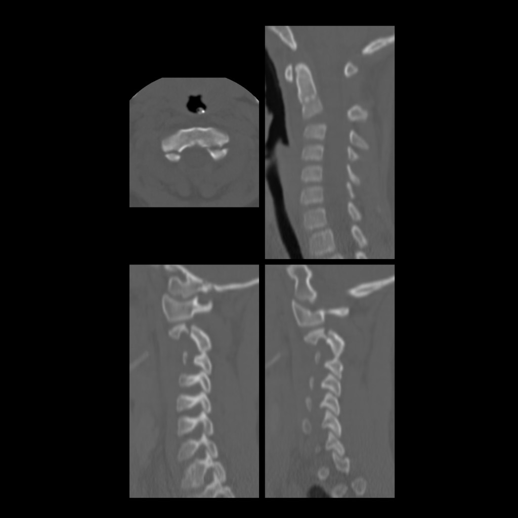
Axial CT without contrast of the cervical spine (top) shows a lucency extending through the left C2 facet and another lucency extending through the right transverse process and involving the right transverse foramen which raised concern for possible right vertebral artery injury which was ruled out on a subsequent CT angiogram of the neck. Sagittal 2D reconstructions from the left, center and right of the cervical spine shows lucencies through the left C2 facet (left image), right transverse process (right image) and anterior inferior aspect of the C2 vertebral body (center 2 images).Axial CT without contrast of the cervical spine (upper left) shows lucencies through both pedicles of the C2 vertebral body. Midline sagittal 2D reconstruction (upper right) shows anterior dislocation of the C2 vertebral body on the C3 vertebral body. Left sagittal (lower left) and right sagittal 2D reconstructions (lower right) again show lucencies through the bilateral C2 pedicles and bilateral anterior perching of the C2 inferior facets on the C3 vertebral body.