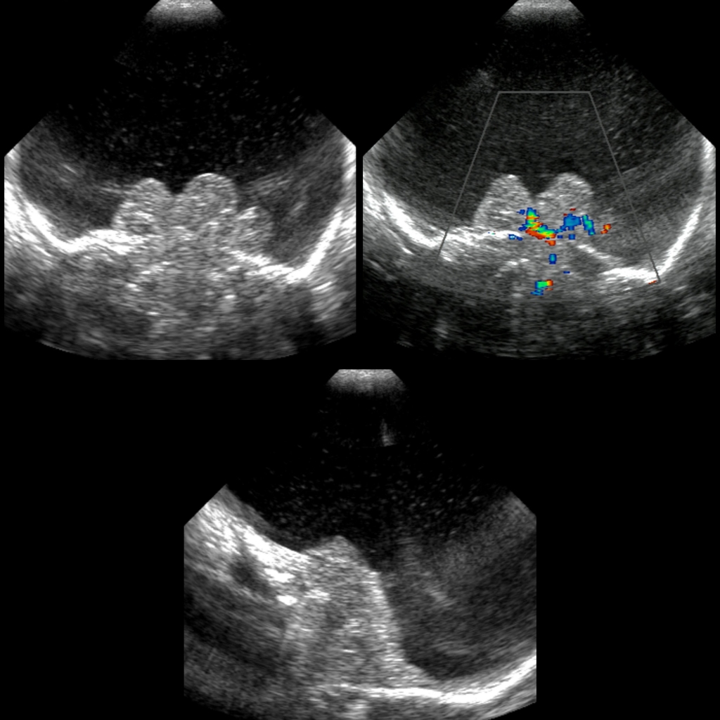
- Etiology: in utero bilateral internal carotid artery occlusion due to in-utero TORCH infection, vascular accident, hypoxic ischemic encephalopathy, maternal carbon monoxide or butane inhalation
- Imaging: have cranial vault, cortical plate and hemispheric white matter destroyed and replaced by thin walled CSF spaces leading to porencephaly of nearly entire cerebral hemisphere, variable presence of thalami and basal ganglia, preservation of brainstem / cerebellum
- DDX: often hard to distinguish from severe hydrocephalus which is possibly treatable
— Differentiating features of hydranencephaly
— Absent MCA/ACA’s with no intact pial vessels / capillary blush
— Usually not macrocephalic
— Focal globular areas of parenchyma in inferior frontal, temporal and occipital lobes
— No diffuse uniform thin gray matter and white matter rim as in hydrocephlus - Complications:
- Treatment:
- Clinical:
Radiology Cases of Hydranencephaly

