Pediatric Hypoxic Ischemic Encephalopathy

  • Etiology: In term infants, 2 types of global hypoxia
    — Partial hypoxia – from decreased cerebral blood flow – affects watershed regions such as periventricular white matter of premature infant (periventricular leukomalacia) or gray matter white matter junction of full term infant, seen best in parasagittal images
    — Profound hypoxia – from cardiac arrest or abruptio placenta – affects regions of highest oxygen demand: basal ganglia, ventral lateral thalami, brainstem, hippocampi, corticospinal tracts, sensorimotor cortex
  • US: Early see diffusely increased parenchymal echogenicity, later see increased echogenicity in basal ganglia
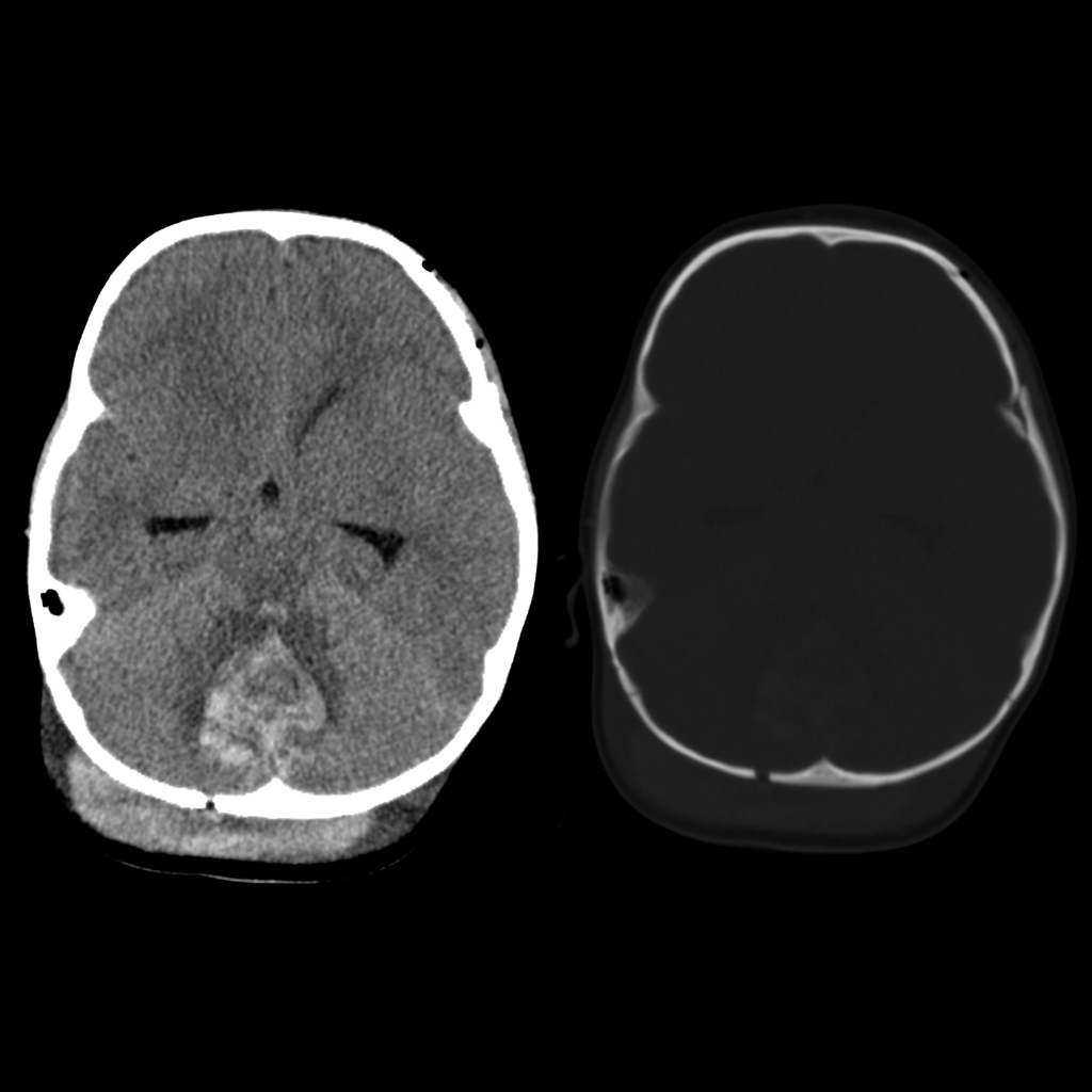
  • CT: Diffuse brain edema with pseudo subarachnoid hemorrhage, white cerebellar sign
  • MRI: Diffuse weighted imaging best performed around 4 days old, in partial hypoxia see peripheral pattern of changes of diffusion restriction at the watershed regions, in profound hypoxia see central pattern of changes of diffusion restriction in the basal ganglia
  • MRI: Appearance depends on severity and duration of the event
    — Moderate and Brief: watershed infarctions
    — Profound and Brief: basal ganglia, thalamus, perirolandic cortex
    — Moderate and Prolonged: diffuse cortex (sparing perirolandic and basal ganglia)
    — Profound and Prolonged: cerebral devastation
  • MRI:
    — In normal infant on T1WI, posterior limb of internal capsule should be bright stripe
    — In profound hypotension on T1WI, loss of signal in posterior limb of internal capsule and increased signal in posterior putamen and ventrolateral thalami indicates bad prognosis
  • DDX:
  • Complications:
  • Treatment: Therapeutic hypothermia for 72 hours with slow and controlled rewarming
  • Clinical: Symptoms are non-specific

  • Hypoxic ischemic encephalopathy in pre term
    — Mild to moderate hypotension – injury to periventricular and deep white matter
    — Profound hypotension: injury to brainstem and thalamus
  • Hypoxic ischemic encephalopathy in full term
    — Mild to moderate hypotension – injury to watershed portions of cerebral cortex and subcortical and periventricular white matter
    — Profound hypotension – injury to brainstem, basal ganglia, thalamus, perirolandic region

Radiology Cases of Hypoxic Ischemic Encephalopathy

Radiology Cases of Neonatal Hypoxic Ischemic Encephalopathy
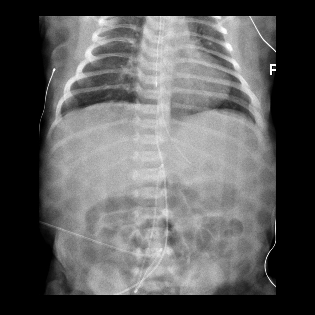
CXR of esophageal temperature probe
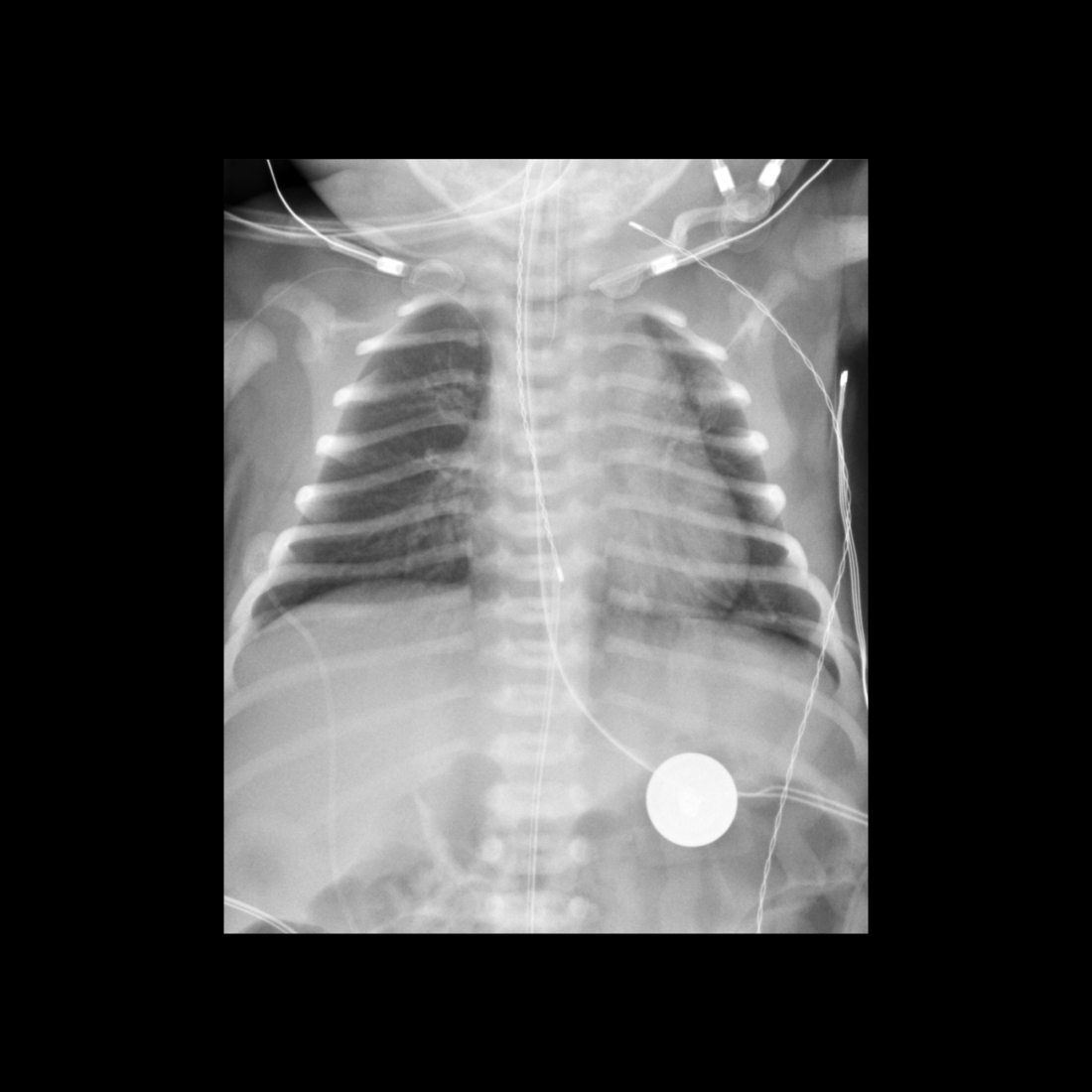
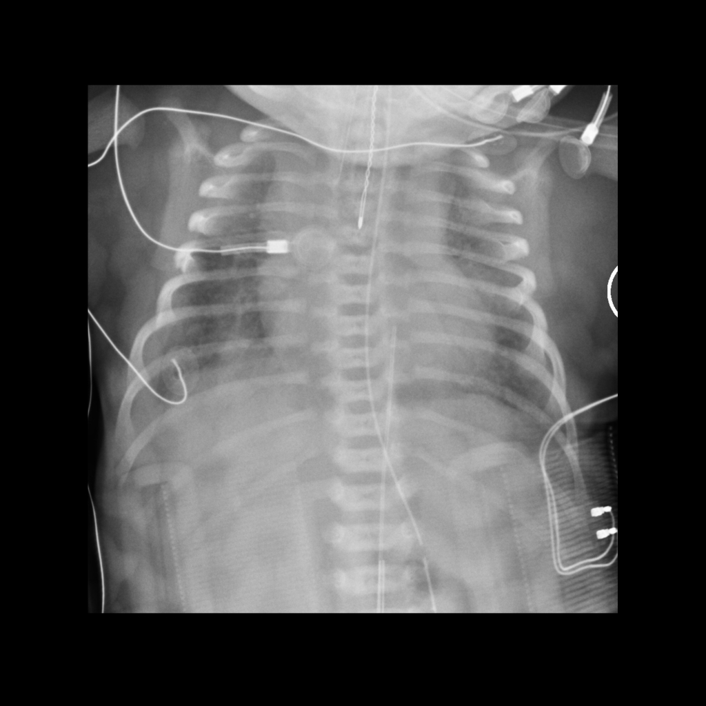
CXR AP shows a nasogastric tube coursing within the esophagus into the stomach while the twisted wire probe next to it with its tip in the mid-esophagus represents an esophageal temperature probe in appropriate position. The patient is lying upon a cooling blanket while being cooled to treat their hypoxic ischemic encephalopathy.
AXR of umbilical arterial catheter in the celiac artery
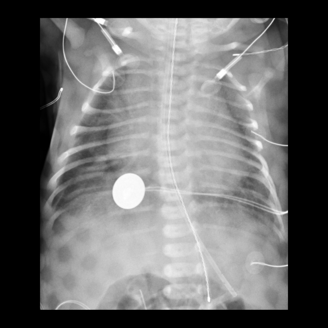
CXR AP shows the tip of the umbilical arterial catheter to be in the celiac artery. The tip of the umbilical venous catheter is at the junction of the inferior vena cava and right atrium. Esophageal temperature probe tip projects over the mid-esophagus. The patient is lying upon a cooling blanket.
CXR of esophageal temperature probe in correct position
CXR AP shows a nasogastric tube coursing in the esophagus with its tip in the body of the stomach. The twisted wire probe coursing next to it with its tip in the mid-to-distal esophagus represents an esophageal temperature probe. Another twisted wire temperature probe is present in the left axilla.
CXR of esophageal temperature probe tip too low in position in the stomach
CXR AP shows a nasogastric tube coursing in the esophagus with its tip in the body of the stomach. The twisted wire probe coursing next to the nasogastric tube with its tip in the body of the stomach represents an esophageal temperature probe. Another twisted wire temperature probe is present in the left axilla.
CT and MRI of hypoxic ischemic encephalopathy
Axial CT without contrast of the brain (above) shows diffuse cerebral edema causing loss of the gray matter white matter junction. Axial T1 without contrast (middle left), FLAIR (middle middle) and T2 (middle right) MRI several days later show developing laminar necrosis. Diffusion weighted imaging (below) shows diffusion restriction throughout the gray matter, white matter, and basal ganglia.

Radiology Cases of Non-Neonatal Hypoxic Ischemic Encephalopathy
CT of caput succedaneum
Axial (above), coronal (below left) and sagittal (below right) CT without contrast of the brain show a large low density fluid collection in the subcutaneous tissues of the scalp that crosses sutures and is seen to surround the skull on the coronal view. Intracranially, there is diffuse loss of gray matter-white matter differentiation secondary to diffuse cerebral edema.
CT of caput succedaneum
Axial CT without contrast of the brain shows a cresenteric high-density fluid collection in the subcutaneous tissues of the right scalp that crosses suture lines and a cresenteric low-density fluid collection in the subcutaneous tissues of the left scalp that crosses suture lines. Intracranially, there is diffuse loss of gray matter-white matter differentiation secondary to diffuse cerebral edema.
CT of hypoxic ischemic encephalopathy and interhemispheric subdural hematoma in child abuse
Axial CT without contrast of the brain shows decreased density of the cerebrum when compared to the density of the cerebellum. There is also loss of gray matter-white matter differentiation and effacement of the cerebral sulci and the basal cisterns. There is linear increased density present along the entire falx.
CT of subgaleal hematoma, diastatic skull fracture, depressed skull fracture, cerebellar contusion, diffuse cerebral edema in child abuse
Axial CT without contrast of the brain shows high density material in the subgaleal tissues posteriorly, a wide lucency in the right posterior skull along with two areas of depressed lucency in the left frontal skull, a rounded high-density lesion in the midline of the cerebellum, and decreased density of the cerebrum when compared to the normal density of the cerebellum along with loss of the normal gray matter-white matter differentiation.
CT of diffuse cerebral edema in child abuse
Axial CT without contrast of the brain shows normal density in the rounded top of the cerebellum (in the center of the left image) compared to the diffuse low-density throughout the cerebrum. There is obliteration of the basal cisterns and loss of the normal gray matter-white matter differentiation. There is also a small left sided high density extra-axial cresenteric fluid collection that tracks medially along the entire falx.