Pediatric Lung Metastasis

  • Etiology: most commonly due to Wilms tumor / rhabdomyosarcoma / Ewing sarcoma / osteosarcoma, less commonly due to lymphoma / neuroblastoma / synovial cell sarcoma / testicular cancer
  • Imaging: usually soft tissue nodule, osteosarcoma metastasis can cause pneumothorax + can be calcified

Radiology Cases of Lung Metastasis

Radiology Cases of Lung Metastasis Due to Wilms Tumor
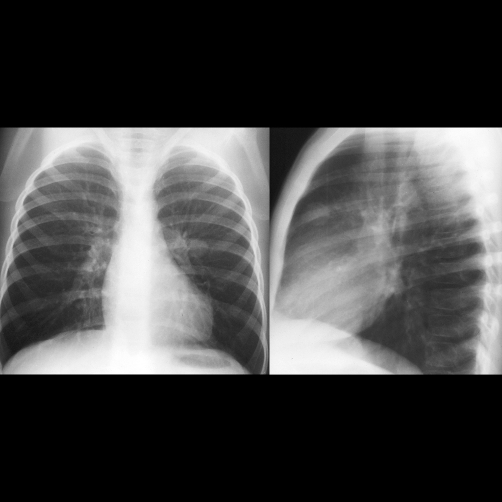
CXR of lung metastasis in Wilms tumor
CXR PA shows a large round opacity just lateral to the left pulmonary artery which is located anteriorly on the lateral view.
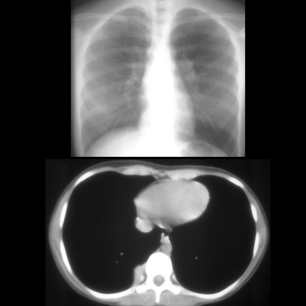
CXR and CT of lung metastasis in Wilms tumor
CXR AP (above) shows a soft tissue density projecting in the right cardiophrenic angle. Axial CT with contrast of the chest (below) shows a soft tissue mass in the right posterior costophrenic sulcus.
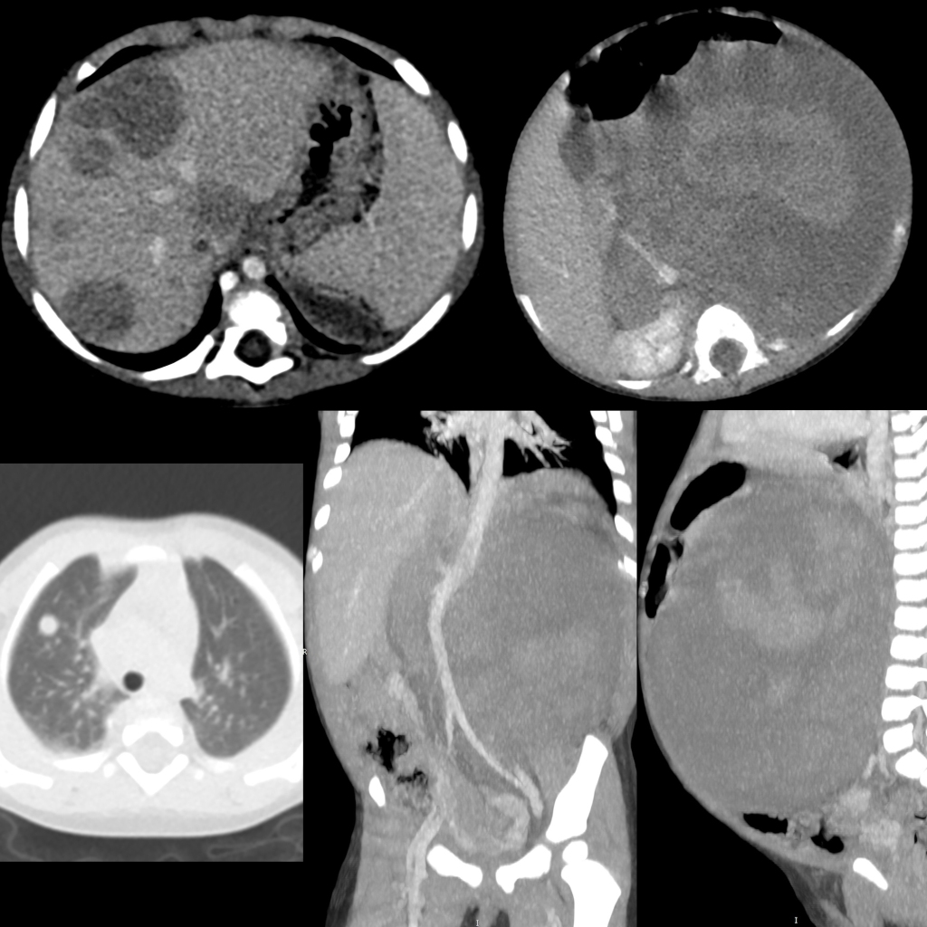
CT of Wilms tumor with lung metastases, liver metastases, and IVC invasion
Axial (above right), coronal (below middle) and sagittal (below right) CT with contrast of the abdomen shows a large heterogenous non-calcified mass that fills the entire left side of the abdomen. The inferior vena cava (to the right of the aorta) was distended with tumor thrombus. Multiple liver (above left) and lung (below left) lesions are also seen.

Radiology Cases of Lung Metastasis Due to Testicle Tumor
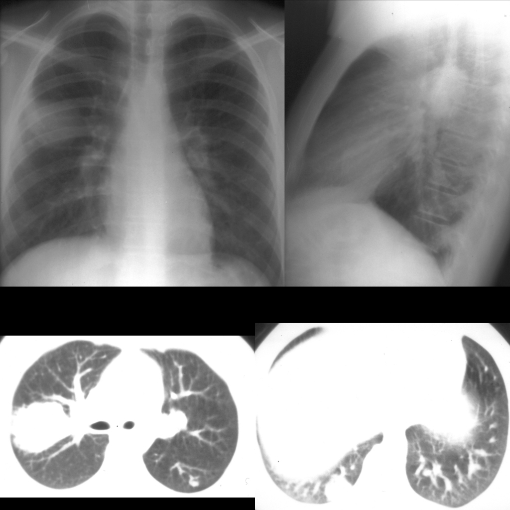
CXR and CT of lung metastases from embryonal cell carcinoma of testicle
CXR PA and lateral (above) shows a large round solid lesion in the right upper lobe. Axial CT without contrast of the chest (below left) shows a large solid lesion in the right upper lobe and a small solid lesion in the left upper lobe posteriorly. There is an additional solid lesion in the right lower lobe (below right).

Radiology Cases of Lung Metastases Due to Fibrolamellar Hepatocellular Carcinoma

CXR of lung metastases in fibrolamellar hepatocellular carcinoma
CXR AP shows multiple large round non-calcified lesions in the lungs bilaterally.