Malrotation With Midgut Volvulus

  • Etiology: prerequisite is malrotation – abnormal rotation of the gut as it returns to the abdominal cavity during embryogenesis -> narrow mesenteric root -> volvulus / twisting of mesenteric pedicle around superior mesenteric artery -> venous obstruction -> continued arterial inflow -> venous congestion -> raised tissue pressure above blood pressure -> cessation of arterial flow / arterial obstruction -> midgut ischemia / necrosis from jejunum to middle of colon
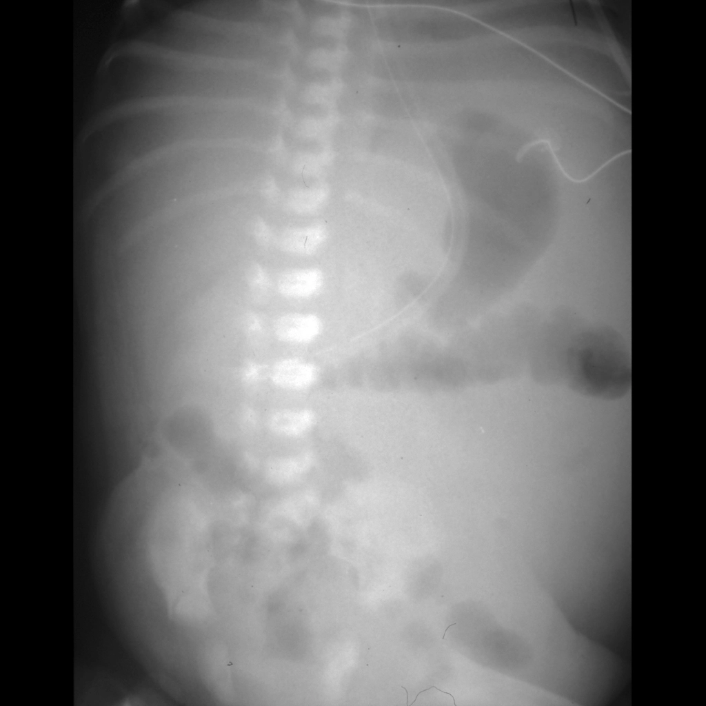
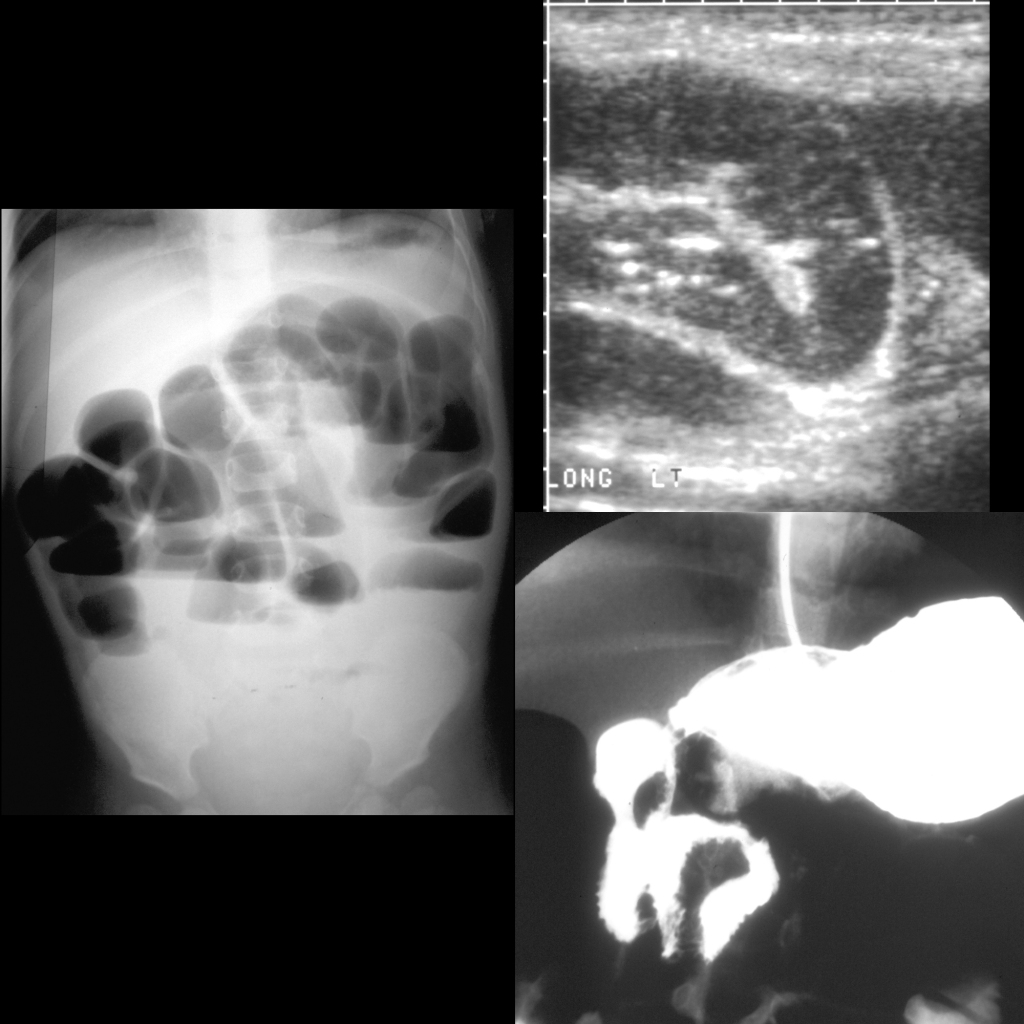
  • AXR: usually unremarkable
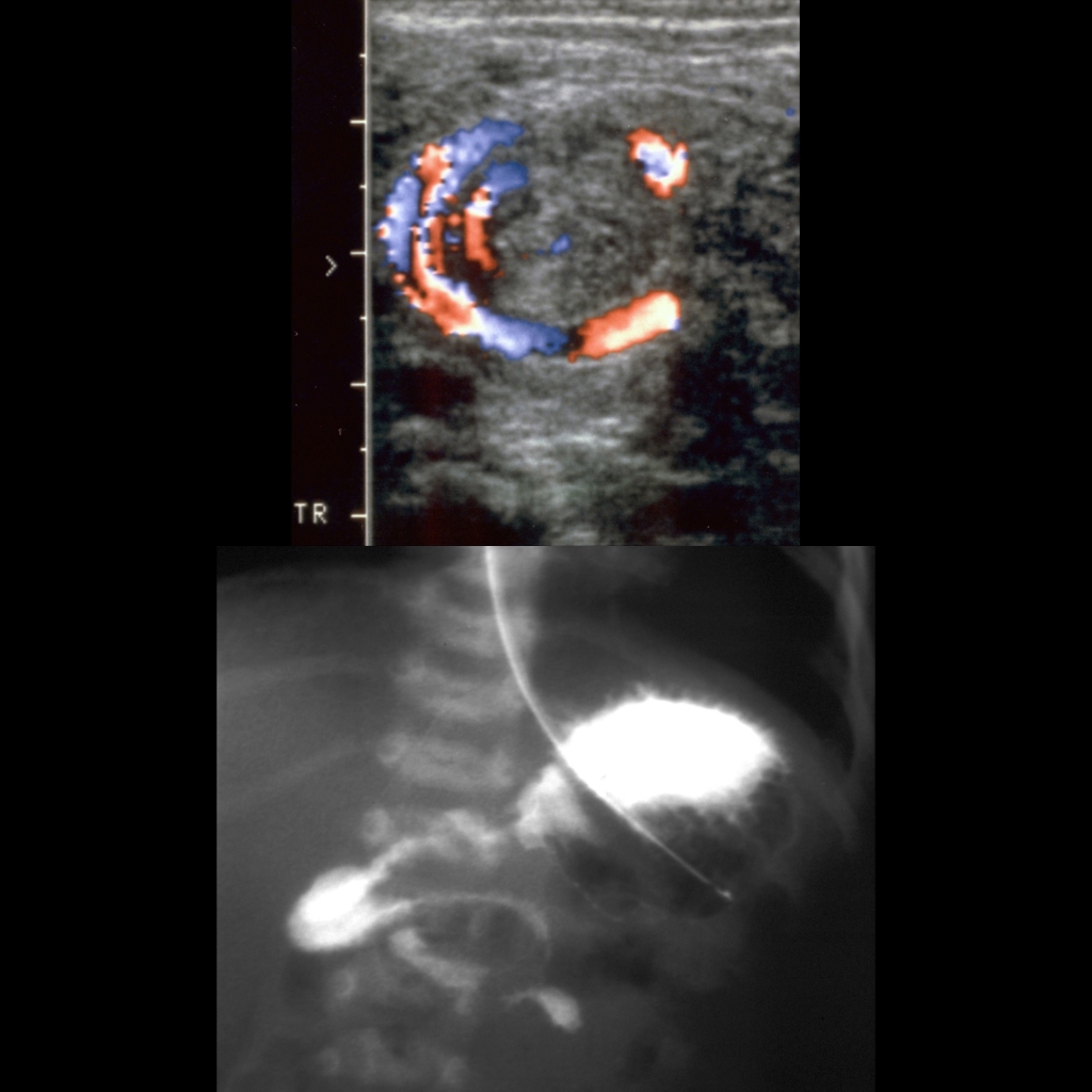
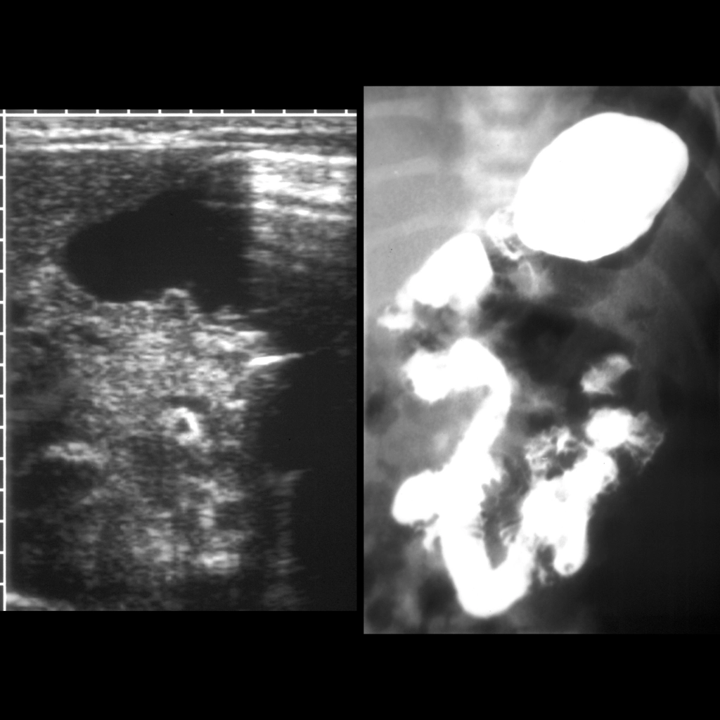
  • US: reversal of normal relationship of superior mesenteric vein and superior mesenteric artery on axial images – super mesenteric vein is to the right of the superior mesenteric artery in the body, if the superior mesenteric vein is above or to the left of the superior mesenteric artery in the body should raise suspicion for malrotation being present, whirlpool sign – swirling of mesenteric vasculature around itself – is sign of midgut volvulus
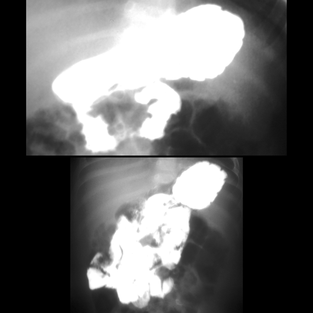
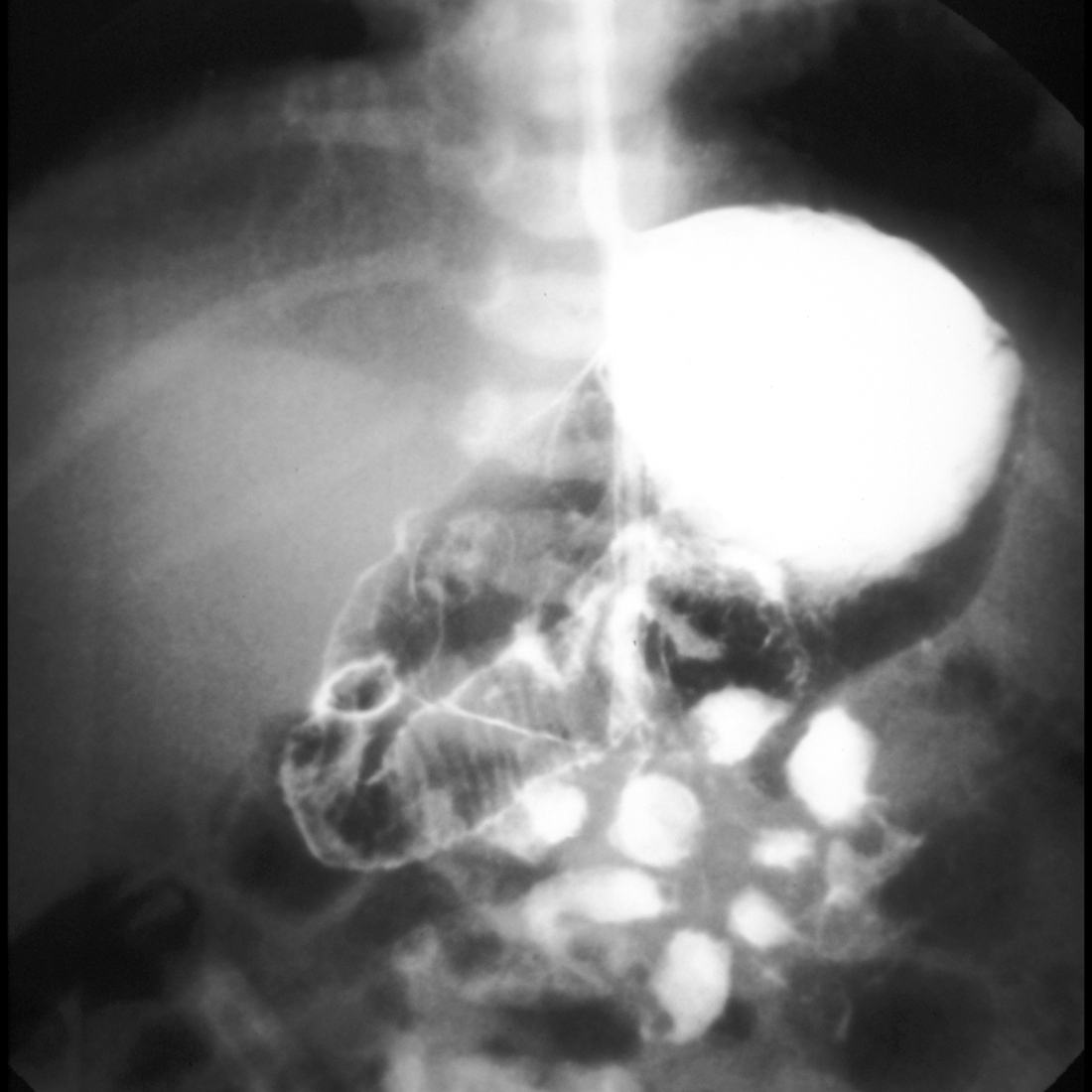
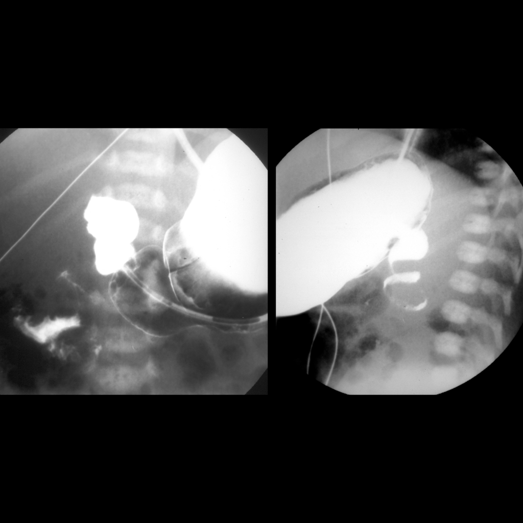
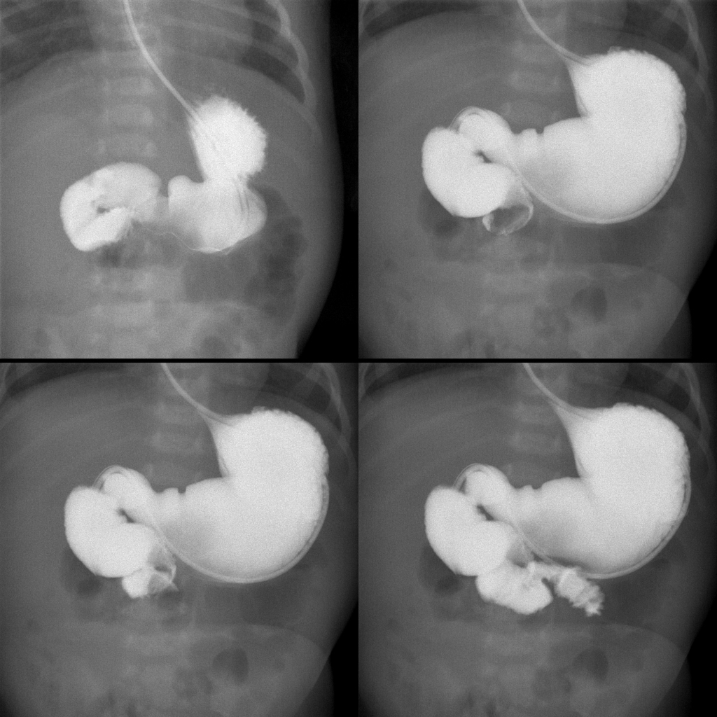
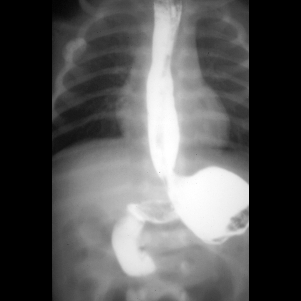
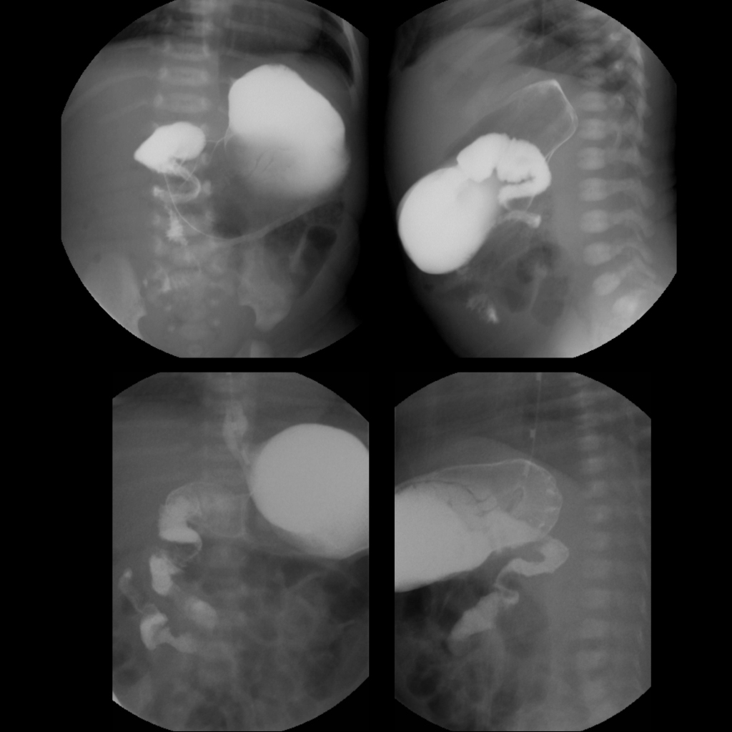
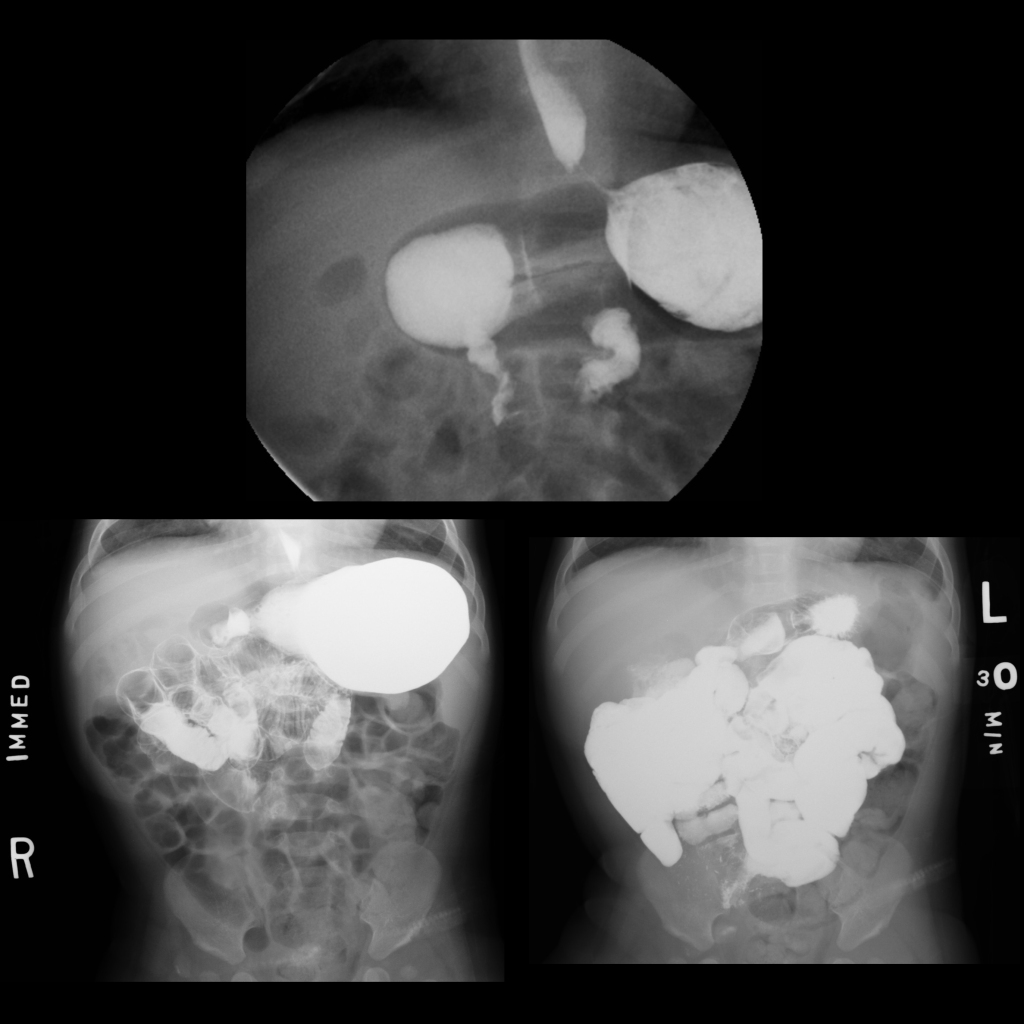
  • UGI: duodenal-jejunal junction (ligament of Treitz) is where retroperitoneal duodenum comes out into peritoneum and it needs to be over left pedicle of spine or to left of spine and to be at level of duodenal bulb and failure to meet both of these criteria results in diagnosis of malrotation with small intestine in right side of abdomen and colon in left side of abdomen, often a spiral / apple peel / corkscrew appearance to the duodenum / proximal jejunum which is a demonstration of the volvulized bowel
  • BE: abnormal position of cecum out of right lower quadrant either high in position in the right abdomen or anywhere on the left side of the abdomen should raise suspicion for malrotation being present, swirling of the terminal ileum is a sign of midgut volvulus
  • Treatment: Ladd’s procedure
  • Clinical: most occur early in life with 40% in first 10 days and 90% in first 3 months of life, bilious vomiting is radiological and surgical emergency as strangulation sequence can happen in few hours and time = bowel, in older children and adults may present as episodes of spontaneously resolving duodenal obstruction

Radiology Cases of Malrotation With Midgut Volvulus

Radiology Cases of Normal Bowel Rotation

UGI of normal rotation of the bowel
AP image from an UGI exam (above) shows the duodenal jejunal junction to project to the left of the spine in its normal position. Later image from the exam (below) shows the jejunum to be on the right side, rather than the left side, of the abdomen.
UGI of normal rotation of the bowel
AP image from an upper GI exam shows a normal appearing duodenal C loop with the duodenal-jejunal junction projecting to the left of the spine at the same level as the duodenal bulb. The jejunum is on the left side of the abdomen.
Upper GI of normal position of ligament of Treitz
AP image from an upper GI exam shows a dilated air and contrast filled stomach and duodenum. The duodenal-jejunal junction is normally positioned at the level of the duodenal bulb and to the left of the spine at L1.

Radiology Cases of Bilious Residuals After Feeding Due to Nasogastric Tube Malposition Clinically Mimicking Malrotation With Midgut Volvulus

AXR of nasogastric tube tip in duodenum
AXR AP shows the tip of the nasogastric tube to be transpyloric in position, in the first part of the duodenum.

Radiology Cases of Malrotation With Midgut Volvulus

AXR of malrotation with midgut volvulus presenting with a double bubble sign
AXR AP shows a very dilated stomach and proximal duodenum (double bubble sign) along with some distal bowel gas.
AXR of small bowel obstruction due to malrotation with midgut volvulus
AXR supine shows multiple dilated loops of bowel with thickened walls.
UGI of malrotation with midgut volvulus
Two AP images from an upper GI exam show the duodenal-jejunal junction to be to the left of the spine but to be low lying. The proximal jejunum has a spiral appearance.
Upper GI of malrotation with midgut volvulus
AP image from an upper GI (left) shows the ligament of Treitz projecting over the right pedicle of the L2 vertebral body and to be lower in position than the duodenal bulb that projects over the right pedicle of the L1 vertebral body. The spiral appearance of the proximal jejunum is best appreciated on the lateral image (right).
UGI of malrotation with midgut volvulus
AP image from an UGI exam (left) shows dilation of the stomach and first and second parts of the duodenum. There is an abrupt transition in bowel contour at the ligament of Treitz which projects low in position and over the middle of the spine. The jejunum then appears to spiral away downstream. AP image from later in the exam (right) better shows the spiraling / corkscrew appearance of the proximal jejunum.
UGI of malrotation with midgut volvulus
AP image from an upper GI exam (left) shows the duodenal jejunal junction to be located to the right of the spine. Lateral image (right) shows the duodenum to have a corkscrew appearance.
UGI of malrotation with midgut volvulus
Serial AP images from an UGI exam show dilation of the first and second parts of the duodenum, the ligament of Treitz projects over the midline of the spine and lower than the duodenal bulb, and the third portion of the duodenum has a spiral or corkscrew appearance.
Upper GI of malrotation with midgut volvulus
AP image from an upper GI shows complete obstruction of the duodenum at the junction of the second and third parts of the duodenum and associated massive gastroesophageal reflux. A final image taken 5 minutes later was unchanged.
UGI of malrotation with midgut volvulus
AP image from an UGI exam shows dilation of the first and second parts of the duodenum. The ligament of Treitz was located in the middle of the spine and was in a lower position than the duodenal bulb. The proximal jejunum was in the right upper quadrant.
Color US of Whirlpool sign of malrotation with midgut volvulus
Transverse color US of the upper abdomen in the midline (above) shows the mesenteric vessels swirling around themselves (Whirlpool sign). The superior mesenteric vein was above the superior mesenteric artery. Upper GI exam (below) shows the duodenal jejunal junction to be to the left of the spine but to be lower in position than the duodenal bulb and the proximal jejunum appears to spiral downwards away from it.
US and UGI of malrotation with midgut volvulus
Transverse US of the pylorus (left) showed a normal appearing pylorus and showed the superior mesenteric vein to be directly above the superior mesenteric artery (the round structure with an echogenic rim in the center of the image), raising suspicion for malrotation. AP image from an UGI exam (right) shows the duodenal jejunal junction to be over the right pedicle of the spine and to be below the level of the duodenal bulb.

Radiology Cases of Malrotation With Intermittent Midgut Volvulus

UGI of malrotation with intermittent midgut volvulus
AP image (above left) obtained early during an upper GI exam shows the duodenal bulb positioned over the spine and the second part of the duodenum to project to the left of the spine and to terminate in a beak with no contrast passing beyond this point. Lateral image (above right) confirms the complete duodenal obstruction. AP image (below) obtained 5 minutes later now shows the obstruction / midgut volvulus to be resolved as the ligament of Treitz is now positioned below the duodenal bulb and to the right of the spine. The proximal jejunum is on the right side of the abdomen.

Radiology Cases of Malrotation With Midgut Volvulus Before and After Ladd Procedure

UGI of malrotation with midgut volvulus before and after a Ladd procedure
AP (above left) and lateral (above right) images from an upper GI exam on day of life 1 show dilation of the first part of the duodenum and a spiral or corkscrew appearance of the second and third parts of the duodenum with the ligament of Trietz projecting over the midline of the spine and lower than the first part of the duodenum. AP (below left) and lateral (below right) images from an upper GI obtained several days after a Ladd procedure show the first part of the duodenum now to be normal in caliber while the second and third parts of the duodenum continue to have a spiral or corkscrew appearance with the ligament of Trietz continuing to project over the midline of the spine and lower than the first part of the duodenum.

Radiology Cases of Malrotation With Chronic Midgut Volvulus

Upper GI of malrotation with chronic midgut volvulus
AP image from a small bowel follow through exam (below) shows the small bowel on the right side of the abdomen and the colon on the left side of the abdomen. Close examination of the upper GI portion of the exam (above) beyond the abnormal position of the ligament of Treitz shows a thickened appearance of the folds throughout the duodenum (above left) and proximal jejunum (above right) but there was no evidence of spiraling of the bowel or obstruction.

Radiology False Positive Cases of Malrotation With Midgut Volvulus

UGI of false positive malrotation with midgut volvulus
Early AP image from an UGI exam (above) showed the ligament of Treitz to project over the midline of the spine and at the same level as the duodenal bulb. Later image (below left) showed the proximal jejunum in the right upper quadrant. Final image (below right) after following contrast through to the colon showed the cecum to be on the right side of the abdomen but to be high in position. The patient was surgically explored and in the operating room the ligament of Treitz was seen in the left upper quadrant and there was normal rotation of the bowel.

Radiology Cases of False Positive Cases of Malrotation With Midgut Volvulus Due to Displacement of the Duodenal-Jejunal Junction by Dilated Loops of Small Bowel

UGI of false positive malrotation with midgut volvulus
AXR upright (left) shows multiple dilated loops of small bowel with air fluid levels. Sagittal US of the abdomen (above right) shows multiple dilated loops of peristalsing small bowel, however an intussusception was not seen. AP image from an upper GI exam (below right) shows the duodenal-jejunal junction to be low in position and to be to the right of the spine.

Surgery Cases of Malrotation With Midgut Volvulus

Surgical image
Surgical image shows the entire small bowel, from the jejunum to the terminal ileum, to be infarcted.
Surgical image of malrotation with midgut volvulus
Surgical image shows the midgut is dusky but not necrotic.
Surgical image of malrotation with midgut volvulus
Surgical image shows the entire midgut being twisted around a single narrow mesentery in the center of the image.
Surgical image of malrotation with midgut volvulus
Surgical image shows spiraling of the bowel around the midgut mesentery in the center of the image.
Surgical image of malrotation with midgut volvulus
Surgical image shows the midgut (superiorly) to be dusky in color.
Surgical image of malrotation with midgut volvulus
Surgical image shows spiraling of the bowel around the midgut mesentery in the center of the image. The small bowel (to the left) is dusky in color.
Surgical image of malrotation with midgut volvulus
Surgical image shows spiraling of the bowel around the midgut mesentery in the center of the image. The small bowel is not dusky in color.
Surgical image of malrotation with midgut volvulus
Surgical image shows spiraling of the bowel around the midgut mesentery in the center of the image. The small bowel (inferiorly) is dusky in color.
Surgical image of malrotation with midgut volvulus
Surgical image shows spiraling of the bowel around the midgut mesentery (right superiorly). The small bowel (peripherally) is dusky in color.
Surgical image of malrotation with midgut volvulus
Surgical image shows spiraling of the bowel around the midgut mesentery (superiorly). The small bowel (peripherally) is dusky in color.
Surgical image of malrotation with midgut volvulus
Surgical image shows spiraling of the bowel around the midgut mesentery (left inferiorly). The small bowel (superiorly) is dusky in color.