Pediatric Tracheobronchial Papillomatosis

  • Etiology: aerosolized during laser resection and spreading from airway to lung
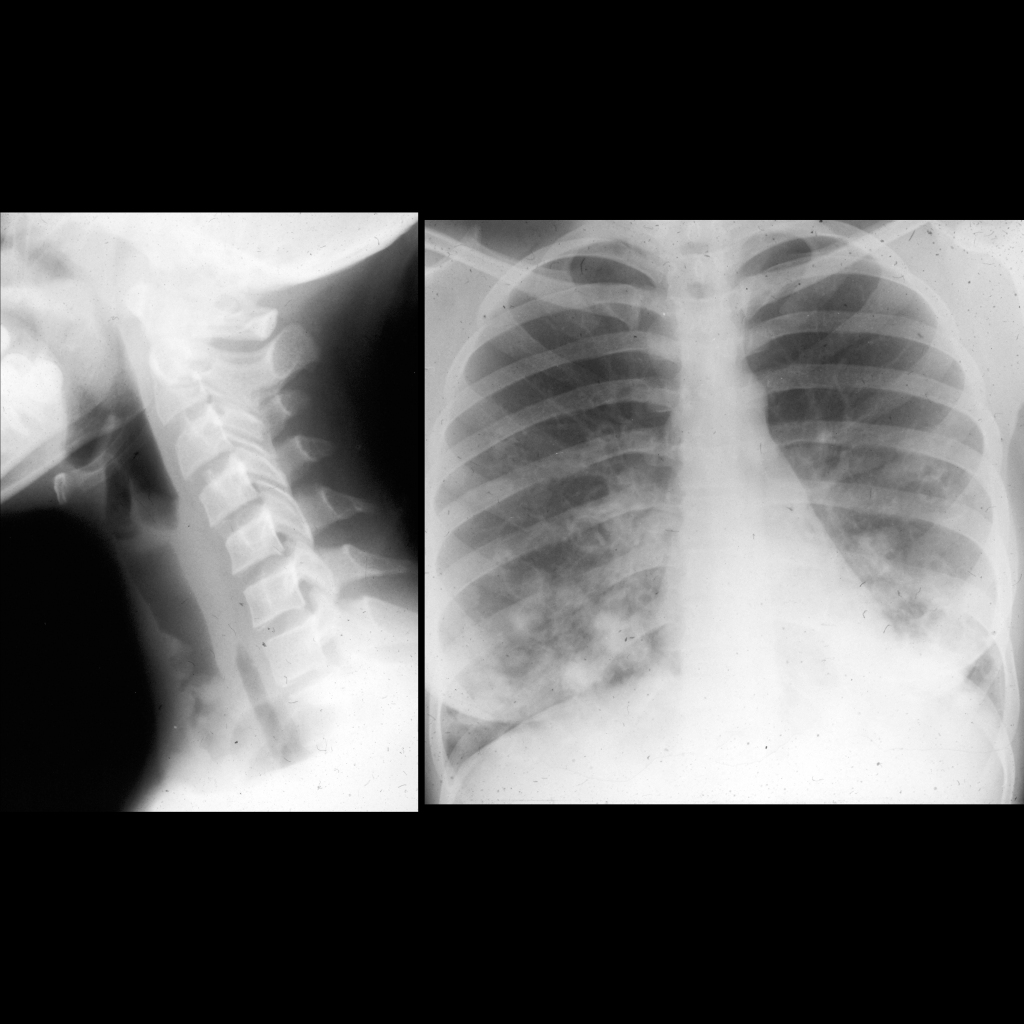
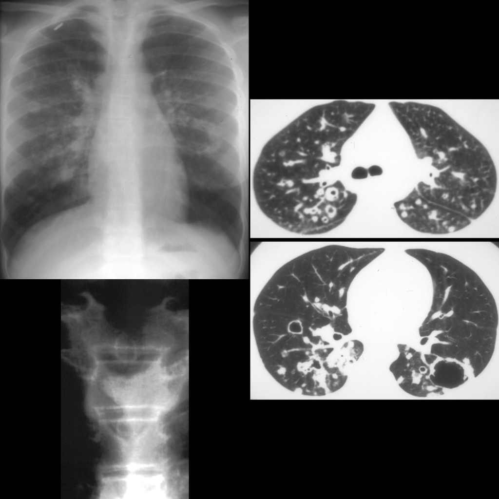
  • Imaging: solid or cystic nodules (thin wall), bronchiectasis, mucous plugs
  • Complications: squamous cell carcinoma of lungs
  • Clinical: chronic cough, hemoptysis, infiltrates

Radiology Cases of Tracheobronchial Papillomatosis

Radiograph and CXR of tracheobronchial papillomatosis
Lateral radiograph of the airway shows multiple polypoid lesions in the trachea. CXR shows multiple cystic lesions in the chest.
CXR and CT of tracheobronchial papillomatosis
CXR AP (above left) shows multiple thin walled cystic and solid lesions in both lungs. Axial CT without contrast of the chest (right) shows multiple cystic and solid lesions in the lungs bilaterally. AP image of the airway (below left) shows a mural tracheal mass arising from the left side of the trachea which nearly completely occludes the airway.

Gross Pathology Cases of Tracheobronchial Papillomatosis

Gross pathology image of papillomas
Pathological image shows multiple white subglottic tracheal masses.