Pediatric Rib Fracture

  • Etiology: blunt thoracic trauma
  • Imaging: spectrum from subtle cortical irregularity of rib contour to frank displacement of fracture fragments, look for underlying pneumothorax / lung parenchymal injury / liver + spleen injury
  • Clinical: unexplained rib fracture in infant or toddler has high specificity for child abuse

Radiology Cases of Rib Fracture

Radiology Cases of Rib Fracture Due to Blunt Chest Trauma

CXR and CT of rib fracture and pulmonary contusion
CXR AP shows cortical irregularities of the right 6th and 7th ribs and no evidence of lung injury. Axial CT of the chest shows a large amount of airspace disease in the right lower lobe and a very small amount of free air anteriorly in the right hemithorax.
CXR and CT of pneumothorax and pulmonary contusion and rib fracture
CXR AP shows a widened mediastinum and a small right-sided pneumothorax. Axial CT with contrast of the chest with lung (above) and bone (below) windows shows air in the pleural space anterior to the right lung and fluid in the pleural space posterior to the right lung as well as airspace disease in the right lower lobe and a cortical disruption through one of the right ribs posteriorly.
CXR of rib fracture and pleural cap
CXR AP shows a discontinuity at the lateral aspect of the left second rib. There is a pleural cap at the apex of the left hemithorax.

Radiology Cases of Rib Fracture Due to Birth Trauma

CXR of posterior rib fracture due to birth trauma
CXR AP shows clear lungs and an area of callus surrounding the right 7th rib posteriorly.
CXR of posterior rib fractures due to birth trauma
CXR AP shows lucent lines without surrounding periosteal reaction through the left 5th-7th ribs posteriorly.

Radiology Cases of Rib Fracture Due to Child Abuse

CXR of posterior rib fractures in child abuse
CXR AP shows poorly expanded but grossly clear lungs bilaterally. There is dense periosteal reaction / callus formation around the posterior aspect of the left 8th and 9th ribs.
CXR of posterior rib fractures in child abuse
CXR AP shows lucent lines without surrounding periosteal reaction through the right 6th-8th and left 6th-11th ribs posteriorly.
3D CT construction of anterior rib fractures in child abuse
3D CT reconstruction with contrast of the chest shows callus formation around the right 4th-8th ribs and left 4th-9th ribs anteriorly.
CXR of posterior rib fractures which were initially missed in child abuse
CXR AP (above) from initial skeletal survey was unremarkable. Repeat CXR AP (below) from follow-up skeletal survey 2 weeks later shows interval development of periosteal reaction around the left 8th-9th ribs posteriorly.
Radiograph of femur fracture in child abuse
AP and lateral radiographs of the right femur (above) show a linear lucency through the diaphysis of the right femur with no associated periosteal reaction. CXR AP shows callus formation around the lateral aspect of the right 5th-6th ribs.
Radiograph of femur fracture in child abuse
AP radiograph of the right femur shows a lucency through the diaphysis of the right femur with no associated periosteal reaction. CXR AP shows callus formation around the right 7th and 9th ribs laterally, the right 7th-9th ribs posteriorly, the left 3rd-7th ribs laterally and the left 8th rib posteriorly.
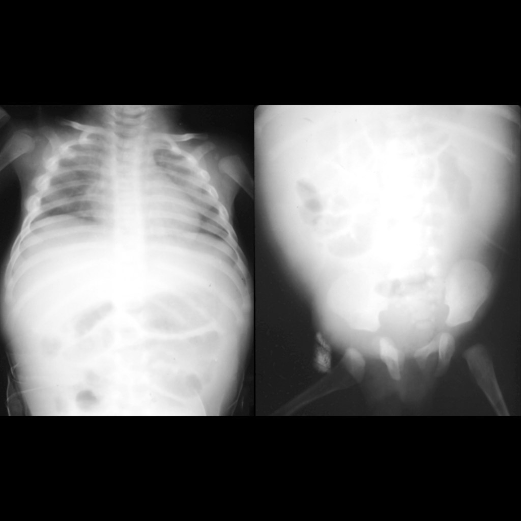
AXR of chylous ascites in child abuse
CXR AP (left) shows the lungs to be poorly expanded and clear but there are dense areas of callus along the lateral aspect of the right 3rd-5th ribs. Centralization of the loops of bowel were also incidentally noted and confirmed on a subsequent AXR AP (right).
CXR of rib fractures in child abuse
CXR AP shows acute fractures of the left 5th through 8th ribs laterally.

Gross Pathology Cases of Rib Fracture

Gross pathological image of posterior rib fractures in child abuse
CXR (above) and complete skeletal survey showed no evidence of rib fractures. Gross pathological image (below) shows callus formation along the posterior aspect of the left 4th-8th ribs.