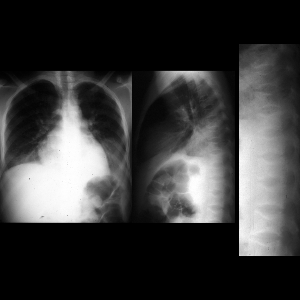
- Etiology:
— Sickling of red blood cells -> anemia -> marrow hyperplasia, marrow reconversion, extra-medullary hematopoiesis
— Vascular occlusion -> bone infarcts, osteonecrosis
— Osteomyelitis due to Staphylococcus aureus + Salmonella - Imaging:
— Marrow hyperplasia: long bones – widening of metaphyses / widening of trabeculae / thinning of cortex / osteopenia, skull – widened diploic spaces
— Bone infarct: in proximal femur / proximal humerus / distal femur / proximal tibia, lucency + sclerosis of medullary bone, cortical bone infarct -> periosteal reaction, H-shaped vertebra due to infarct of central part of vertebral body
— Infection: see erosions / lysis / periosteal reaction, infarction vs. infection appear similar on MR as marrow edema and extra-osseous fluid collections but infarction has high signal intensity on T1WI with fat saturdation due to packed RBCs - Clinical: osteomyelitis is 50 times less common than osteonecrosis, suspect osteomyelitis if patient does not respond to conservative management / has asplenia or impaired complement and phagocytic activity
- Spine T1 marrow changes
— Age < 1 year – vertebral body signal decreased than adjacent disc — 1 year – 5 years – vertebral body signal = adjacent disc — > 5 years – vertebral body signal greater than adjacent disc - Extremity T1 marrow changes
— There is fat in epiphysis within 6 months of radiographic appearance
— Fatty marrow changes in diaphysis of humerus and femur begin by 1 year and complete by 10 years - Normal T1 marrow changes
— During 1st and 2nd decade of life red marrow gradually converts to yellow (fatty) marrow
— In each extremity, conversion occurs from periphery (fingers and toes) to center of body (shoulders and hips)
— In each bone, 1st epiphysis, 2nd diaphysis, 3rd metaphysis - Marrow reconversion with anemia
— Order of transformation back to red marrow is reversed
— Epiphysis are the last part to reconvert
Radiology Cases of Sickle Cell Disease Musculoskeletal


