- Etiology: risk factors – hypercoaguability, contraceptives, dehydration, collagen vascular disease, lumbar puncture
- Imaging: cortical vein thrombosis – thrombus forms central to peripheral, deep dural sinus thrombosis gets bithalamic edema / hemorrhage
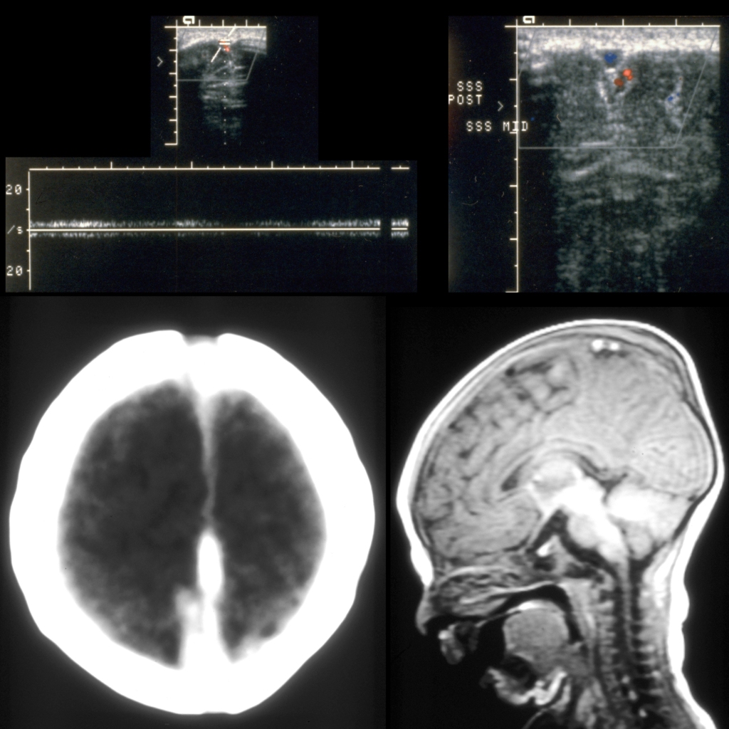
- CT: cord sign – hyperdense cord on non-contrast CT along thrombosed dural sinus
- MRI: T1 hyperintense thrombus in superior sagittal sinus with empty delta on contrast enhanced
- DDX:
- Complications in neonate: venous infarction within region of brain that drains into sinus, involves thalami in medullary vein / straight sinus and Vein of Galen thrombosis, involves cortex and subcortical white matter with sagittal sinus thrombosis, infarction characteristically bilateral and hemorrhagic typically involving the cortex and subcortical white matter
- Treatment:
- Clinical:
Radiology Cases of Dural Venous Sinus Thrombosis
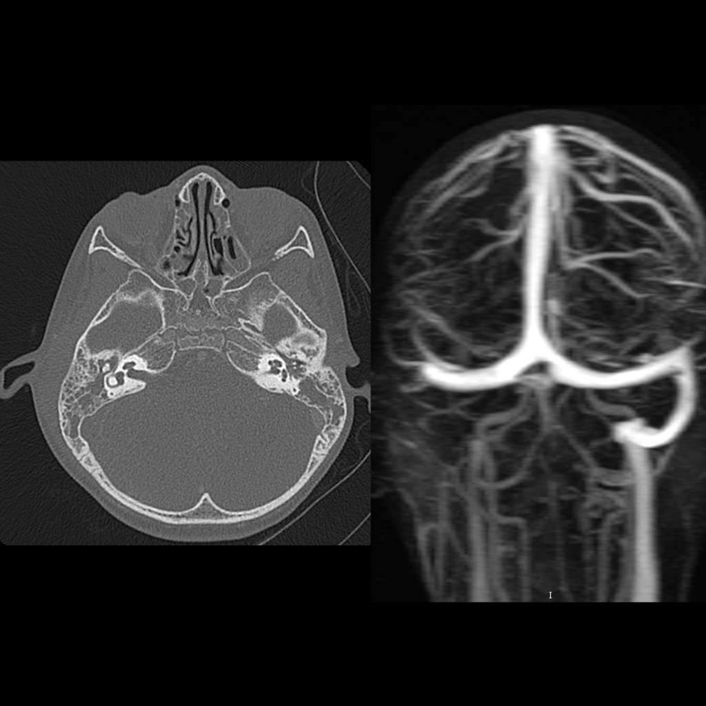
Radiology Cases of Dural Venous Sinus Thrombosis of the Sigmoid Sinus
Radiology Cases of Dural Venous Sinus Thrombosis of the Superior Sagittal Sinus