Pediatric Mediastinal Teratoma

  • Etiology: contains endoderm, mesoderm, ectoderm tissue and therefore classically can contain fat and calcium
  • Layers and what comes from them
    — Ectoderm – neural, epidermis
    — Mesoderm – bone, muscle, connective tissue, dermis, cardiovascular system, reproductive system, kidneys
    — Endoderm – epithelium gastroinstestinal / respiratory / genitourinary, endocrine glands
  • Imaging: anterior mediastinal mass arising from thymus which is often heterogenous and may contain fat and calcium

Radiology Cases of Mediastinal Teratoma

CT of mediastinal teratoma
Axial CT without contrast of the chest shows an anterior mediastinal mass containing soft tissue and fat.
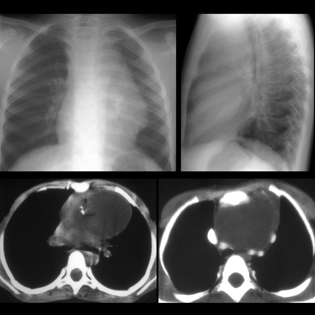
CXR and CT of teratoma of thymus
CXR AP+lateral shows a large anterior mediastinal mass. Axial CT with contrast of the chest shows a heterogenous mass containing low density fat and high density calcium.
CT of mediastinal teratoma
CXR (above left) shows a mass in the left hemithorax. Coronal (above right), axial (below left) and sagittal (below right) CT with contrast of the chest show a large mass of soft tissue and fat density in the anterior mediastinum that compresses the left lung posteriorly and causes mediastinal shift to the right.

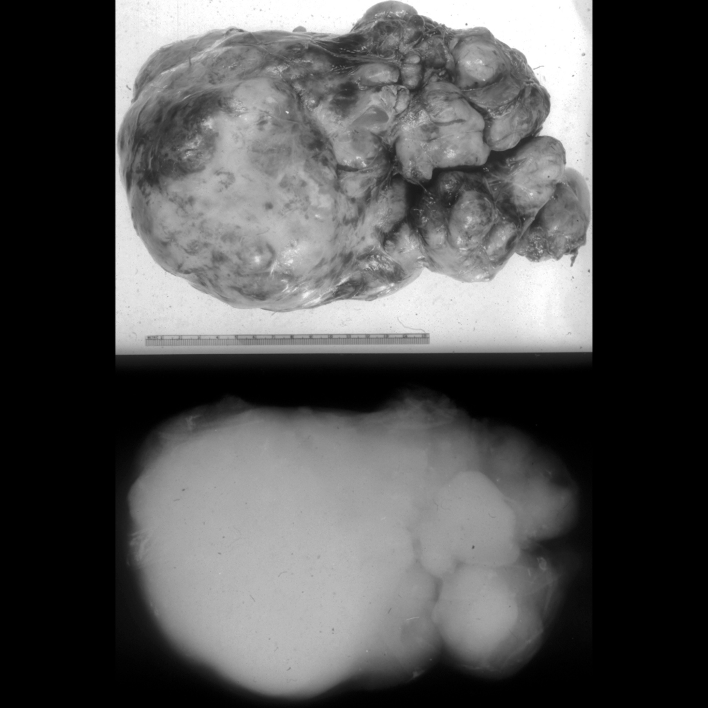
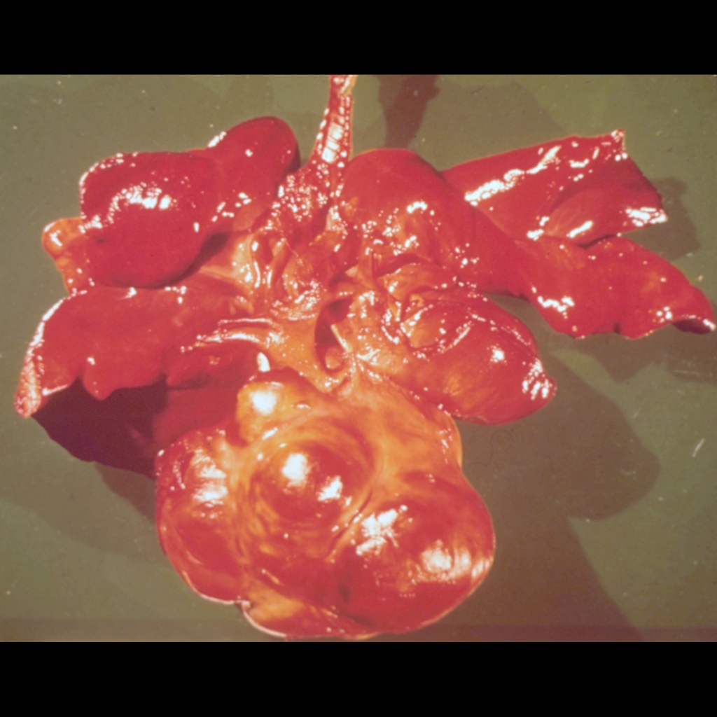
Gross Pathology Cases of Mediastinal Teratoma
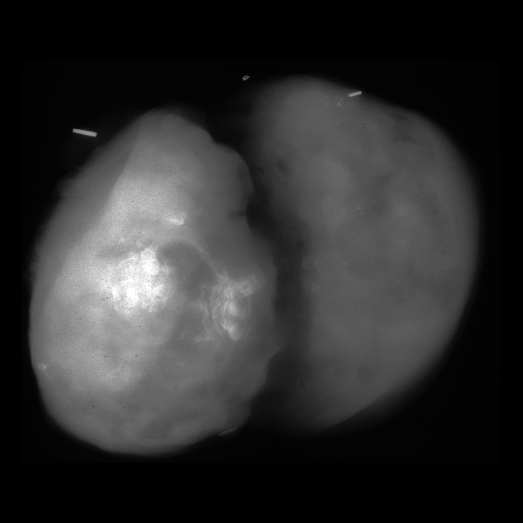
Specimen radiograph of thymic teratoma / mediastinal teratoma
Specimen radiograph shows densely calcified rectangular structures in the resected thymic mass which turned out to be teeth.