- Etiology: Compression of brachial plexus or subclavian vessels as they pass through superior thoracic aperture
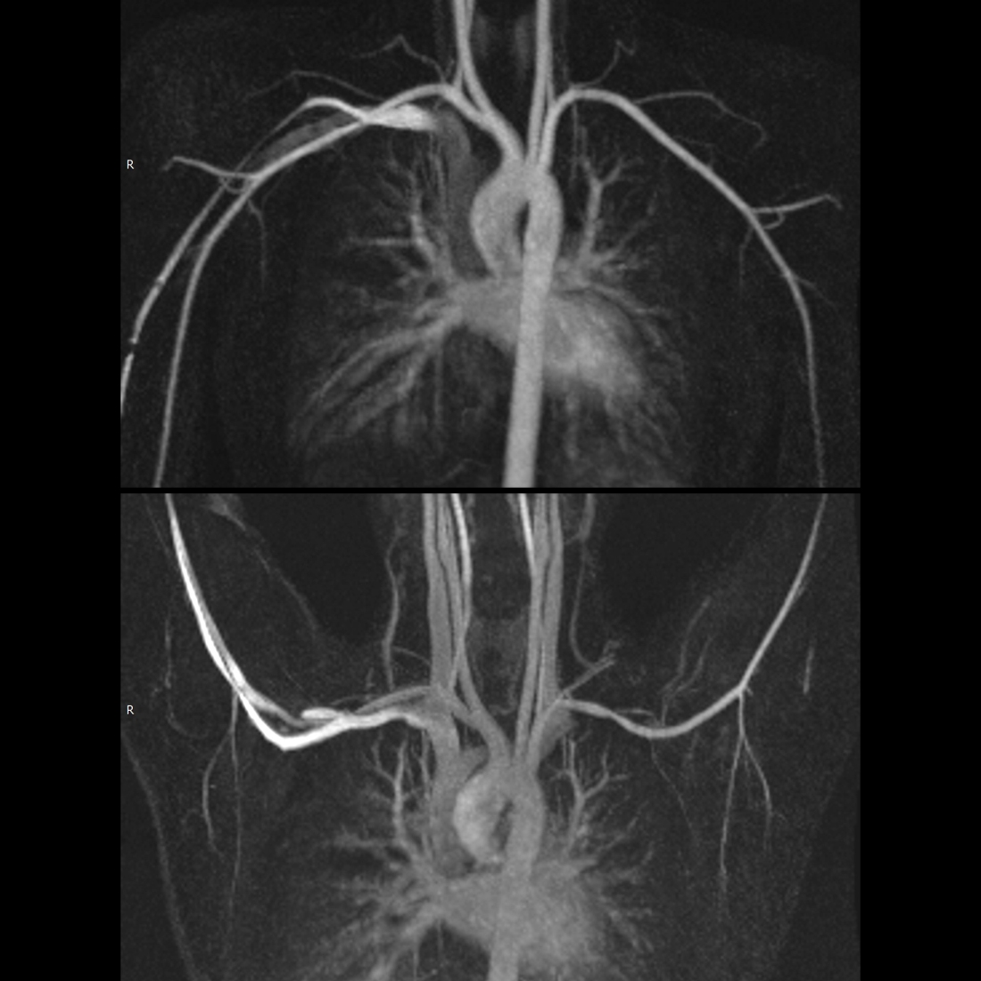
- Imaging: Performed with arms in raised (abducted) and neutral (adducted) positions
— Neurogenic thoracic outlet syndrome: Bone and soft tissue abnormalities, loss of fat around brachial plexus with abduction, edema in brachial plexus
— Venous thoracic outlet syndrome: Bone and soft tissue abnormalities, axillosubclavian vein narrowing with abduction, enlarged collaterals, axillosubclavian vein thrombosis
— Arterial thoracic outlet syndrome: Bone and soft tissue abnormalities, axillosubclavian artery narrowing with abduction, enlarged collaterals, axillosubclavian artery aneurysm or pseudoaneurysm, axillosubclavian artery thrombosis - DDX:
- Complications: Thrombosis of subclavian artery and vein
- Treatment: Surgical
- Clinical:
— Neurogenic thoracic outlet syndrome: Presents with pain / paresthesia / numbness of upper extremity due to brachial plexus compression
— Venous thoracic outlet syndrome: Upper limb swelling and pain due to subclavian vein compression
— Arterial thoracic outlet syndrome: Upper limb ischemia with coolness / pallor / paresthesias / decreased pulses due to subclavian artery compression
Radiology Cases of Thoracic Outlet Syndrome
Radiology Cases of Arterial Thoracic Outlet Syndrome