Tracheal Atresia

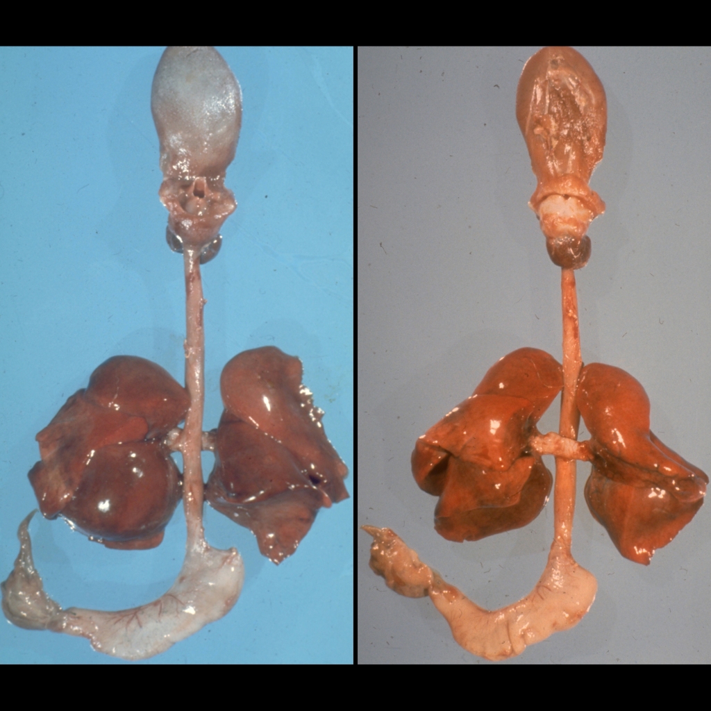
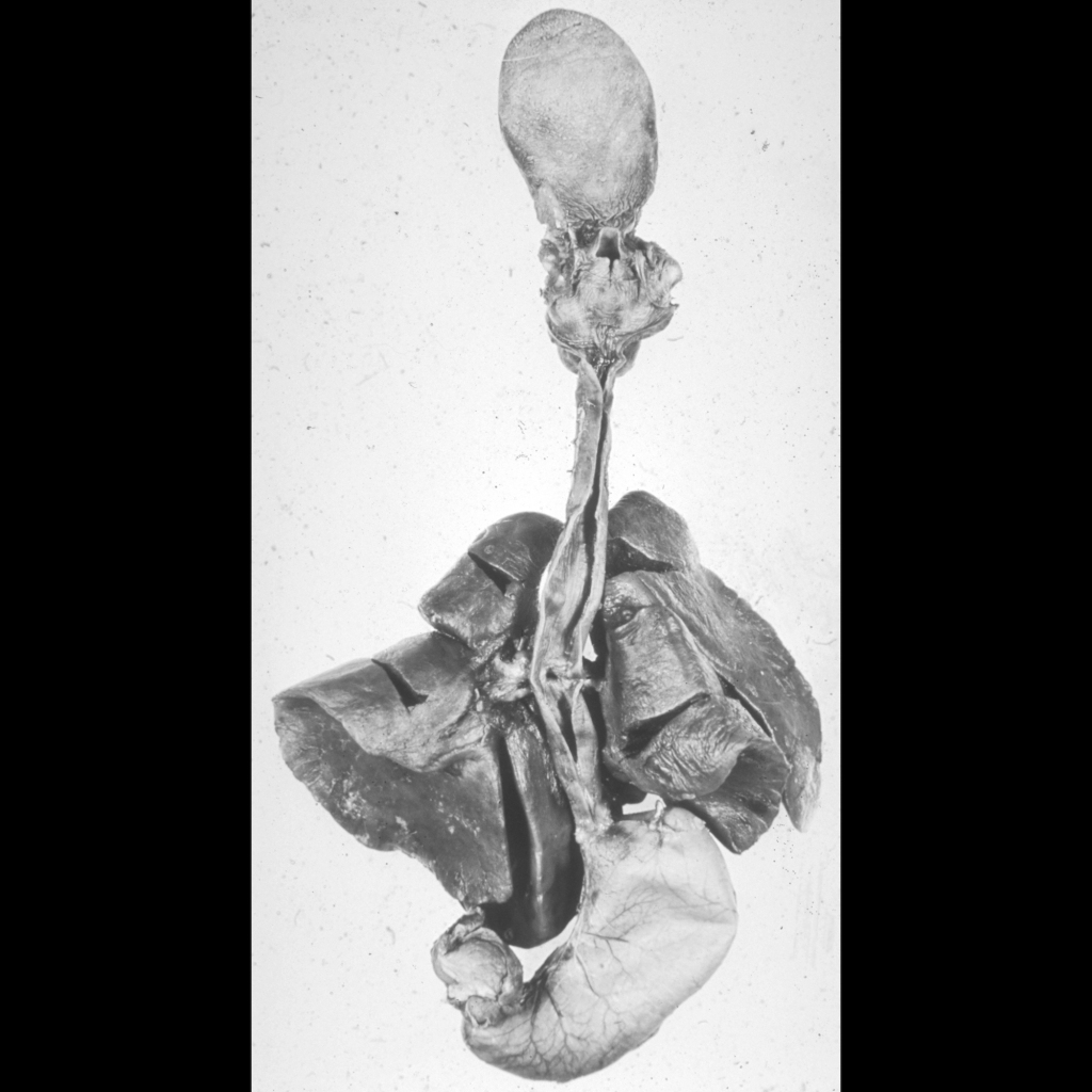
  • Etiology: congenital absence of trachea
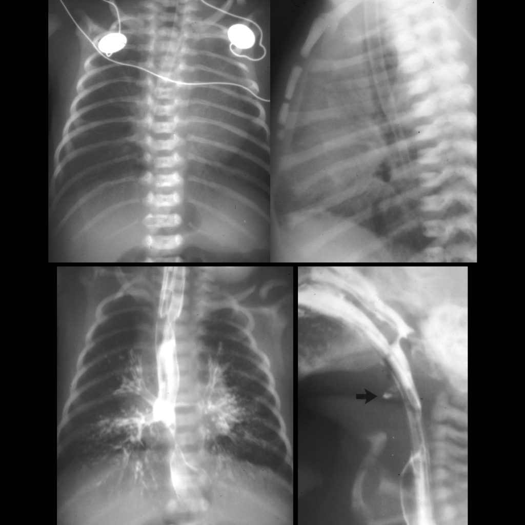
  • Imaging: absence of the trachea, communication between the esophagus and the airways
  • DDX: laryngeal atresia, severe congenital tracheal stenosis
  • Complications: aspiration
  • Treatment:
  • Clinical: in the spectrum of congenital high airways obstruction syndrome (CHAOS)

Radiology Cases of Tracheal Atresia

CXR and UGI of tracheal atresia
CXR AP and lateral (above) shows a nasogastric tube with its tip in the mid-esophagus. AP image from an UGI exam performed by injecting the nasogastic tube (below left) shows simultaneous opacification of the tracheobroncial tree and esophagus. Lateral image from the UGI exam (below right) shows the atretic origin of the trachea (black arrow). The tip of the endotracheal tube (which is in the esophagus) is at approximately the same level.

Gross Pathology Cases of Tracheal Atresia

Pathology image of tracheal atresia
Post-mortem gross pathological images in anterior (left) and posterior (right) projections show absence of the trachea with the mainstem bronchi communicating with the esophagus.
Pathology image of tracheal atresia
Post-mortem gross pathological image with the esophagus sectioned open shows absence of the trachea with the mainstem bronchi communicating with the esophagus as demonstrated in the center of the image.