Umbilical Arterial Catheter Malfunction / Malposition / Misposition / Misplacement

  • Etiology: placed in premature newborns and term newborns who need central arterial access for blood pressure monitoring + blood gas
  • Imaging: normal umbilical artery catheter (UAC) course is umbilical artery to internal iliac artery to common illiac artery to aorta (in and caudad and then cephalad on an AP image) while coursing anterior to the spine on the lateral image, catheter tip should be at T6-T9 vertebral body above celiac artery / superior mesenteric artery / renal arteries (high UAC) or at L3-L5 vertebral body below the inferior mesenteric artery (low UAC), usually projects over left side of spine on AP view
  • Complications: catheter tip positioned above T6 is near origin of great vessels from aortic arch or catheter tip positioned between T9-L2 is near origins of renal arteries and mesenteric vasculature, catheter tip is too low in position, catheter tip thrombosis which can extend into any adjacent arterial vessel and lead to renal infarct / renal artery stenosis / renovascular hypertension / necrotizing enterocolitis / lower extremity ischemia, spinal cord infarction (due to artery of Adamkiewicz) / pulmonary embolus if catheter is placed across a patent ductus arteriosus, perforation of vessel during catheter placement should be suspected when the umbilical arterial catheter projects over no known arterial vessel
  • Treatment: repositioning or removal of malpositioned catheter

Radiology Cases of Umbilical Arterial Catheter Malfunction / Malposition / Misposition / Misplacement

Radiology Cases of Umbilical Arterial Catheter Tip in Appropriate Position

AXR of normal position of an umbilical arterial catheter
AXR AP and cross-table lateral shows normal course of the umbilical arterial catheter from umbilical artery to internal iliac artery to common illiac artery to aorta (in and caudad and then cephalad on the AP view) while coursing anterior to the spine on the lateral view with the catheter tip appropriately positioned at T7. The umbilical venous catheter has a normal course from umbilical vein to left portal vein to ductus venosus to left hepatic vein to inferior vena cava (in and cephalad on the AP view) while coursing through the liver on the lateral view with the catheter tip positioned in the right atrium

Radiology Cases of Umbilical Arterial Catheter Tip Positioned Above T6

CXR of nasogastric tube in mid-esophagus
CXR AP shows the tip of the nasogastric tube in the mid esophagus. The tip of the umbilical venous catheter is too high in the right atrium and the tip of the umbilical arterial catheter is too high in the aortic arch.
CXR of umbilical arterial catheter malposition
CXR AP shows the tip of the umbilical arterial catheter to be at T3 near the aortic arch. The tip of the umbilical venous catheter is in the right portal vein. The lungs show diffuse ground-glass opacity.
CXR of umbilical arterial catheter in left subclavian artery
CXR AP shows the tip of the umbilical arterial catheter to be in the left subclavian artery. The tip of the umbilical venous catheter is deep within the right atrium. The lungs show minimal ground-glass opacity.
CXR of umbilical venous catheter in a pulmonary vein
CXR AP shows the umbilical venous catheter coursing into the right atrium, through a patent foramen ovale, into the left atrium and then into a left pulmonary vein. The umbilical arterial catheter tip is at T4. There is faint ground glass opacity in the lungs.
AXR of umbilical arterial catheter malposition
Supine AXR shows the tip of one umbilical arterial catheter to be at T5 and the tip of the other umbilical arterial catheter to be at T12. Note on this AP image how after insertion at the umbilicus the umbilical arterial catheters first head caudad in the umbilical artery and internal iliac artery before heading cephalad in the common iliac artery and aorta.
AXR of umbilical venous catheter in the right portal vein
AXR AP (above) and cross-table lateral AXR (below) shows the tip of the umbilical venous catheter to be positioned deep within the right portal vein. The tip of the umbilical arterial catheter is at T4.

Radiology Cases of Umbilical Arterial Catheter Tip Between T9-L2
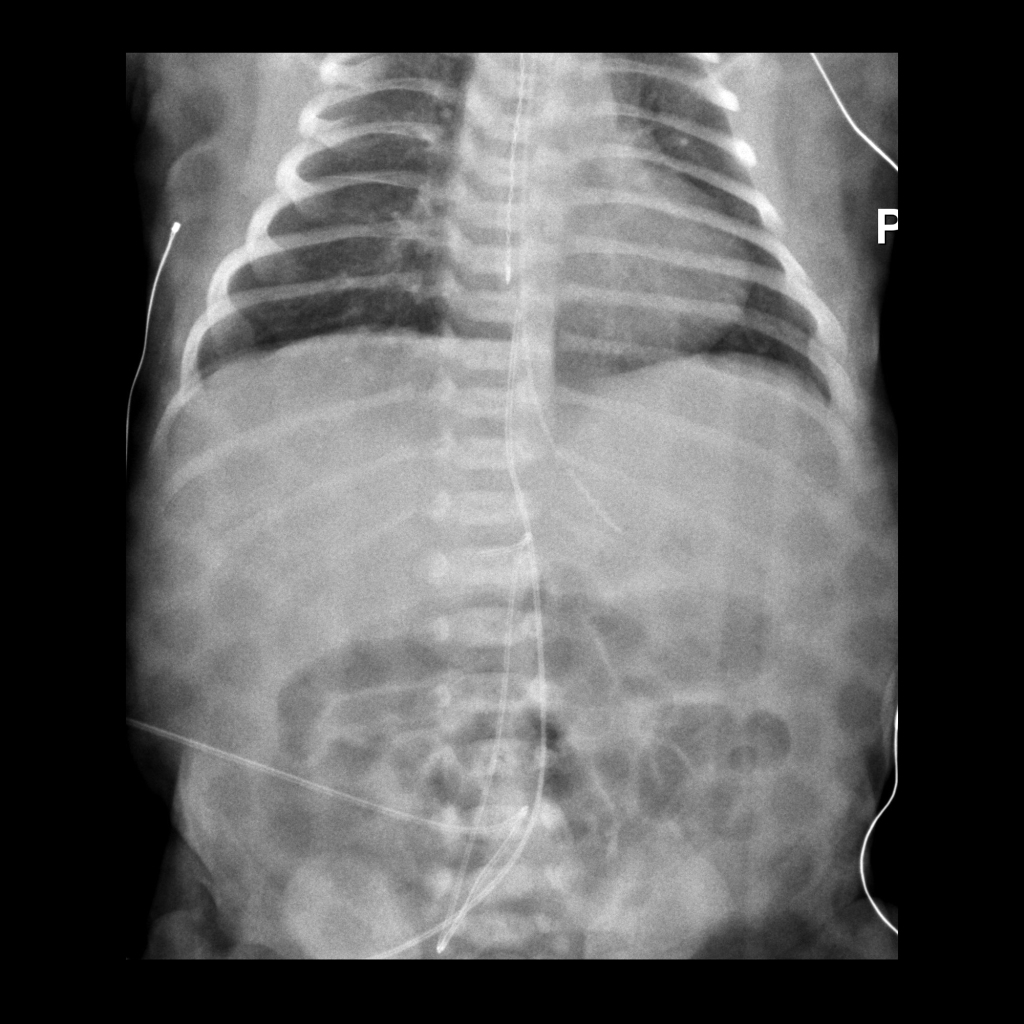
AXR of umbilical arterial catheter in the celiac artery
CXR AP shows the tip of the umbilical arterial catheter to be in the celiac artery. The tip of the umbilical venous catheter is at the junction of the inferior vena cava and right atrium. Esophageal temperature probe tip projects over the mid-esophagus. The patient is lying upon a cooling blanket.
AXR of umbilical arterial catheter malposition
Supine and cross-table lateral AXR shows the tip of one umbilical arterial catheter to be at T6 and the tip of the other umbilical arterial catheter to be at L1. Note on these images how after insertion at the umbilicus the umbilical arterial catheters first head caudad in the umbilical artery and internal iliac artery before heading cephalad in the common iliac artery and aorta. There is faint ground-glass opacity in the lungs.

Radiology Cases of Umbilical Arterial Catheter Tip Below L3

AXR of low malposition of an umbilical arterial catheter
AXR supine shows an umbilical arterial catheter that loops back upon itself in the aorta and whose tip projects in the right iliac artery at S3. The umbilical venous catheter tip projects at the junction of the inferior vena cava and the right atrium.
Radiograph of low-lying umbilical arterial catheter
CXR shows the tip of the umbilical arterial catheter to be at the level of the L2 vertebral body.

Radiology Cases of Umbilical Arterial Catheter Perforation of Vessel

AXR of umbilical arterial catheter perforation
Supine AXR shows the tip of the umbilical arterial catheter to project in the left abdomen over no known arterial vessel. The tip of the umbilical venous catheter projects within the liver.

Radiology Cases of Umbilical Arterial Catheter Tip Thrombus

US of aortic thrombus due to umbilical arterial catheter
Sagittal and transverse grayscale US of the aorta shows an echogenic, linear object within the aorta which on color doppler US was seen to have blood flow around it.
US of thrombus in aorta caused by umbilical arterial catheter
Coronal grayscale US of the aorta centered at the level of the kidneys (above) shows a round echogenic object within the aorta at the level of the origin of the renal arteries. Coronal color doppler US of the aorta (below) shows good blood flow around the object and into the renal arteries which was confirmed on spectral doppler US.