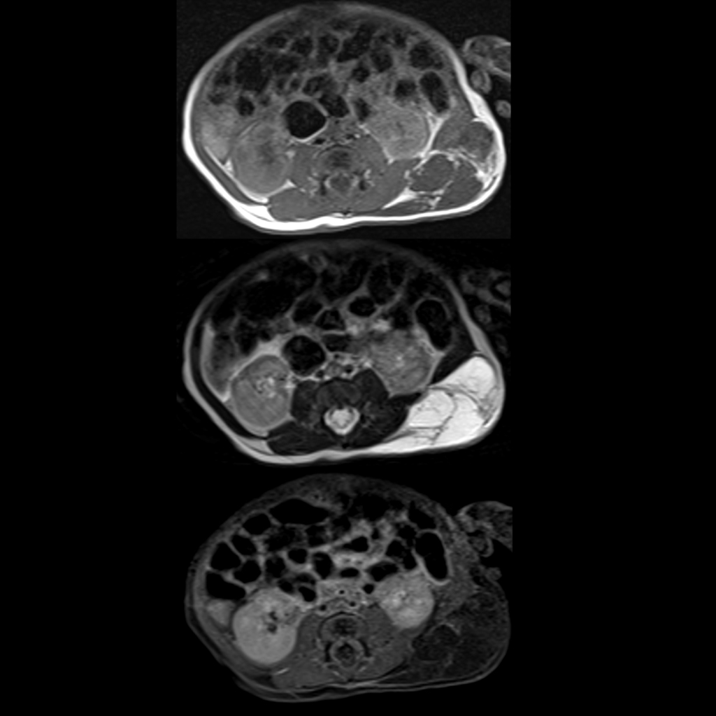
Newborn with a posterior left chest wall mass on prenatal ultrasound Axial T1 without contrast (top), T2 (middle) and T1 with contrast (bottom) MRI of the chest shows a mass composed of multiple large fluid-filled structures separated by thin septations which faintly enhance. The diagnosis was lymphatic malformation.