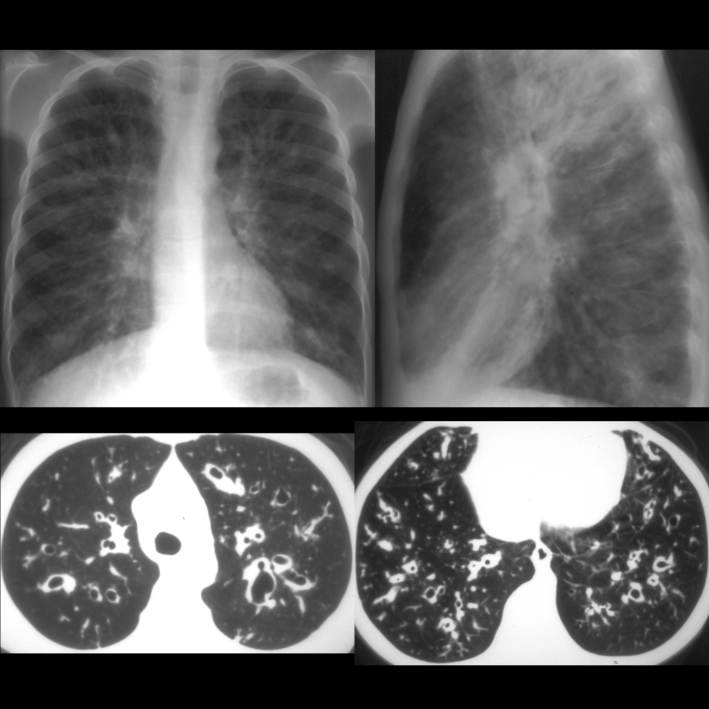
CXR AP and lateral (above) show bilaterally hyperexpanded lungs with a coarse interstitial pattern, bronchial wall thickening and bronchiectasis. Axial CT without contrast of the chest show thickened interlobular septa and bronchiectasis in the bilateral upper (below left) and lower (below right) lobes.