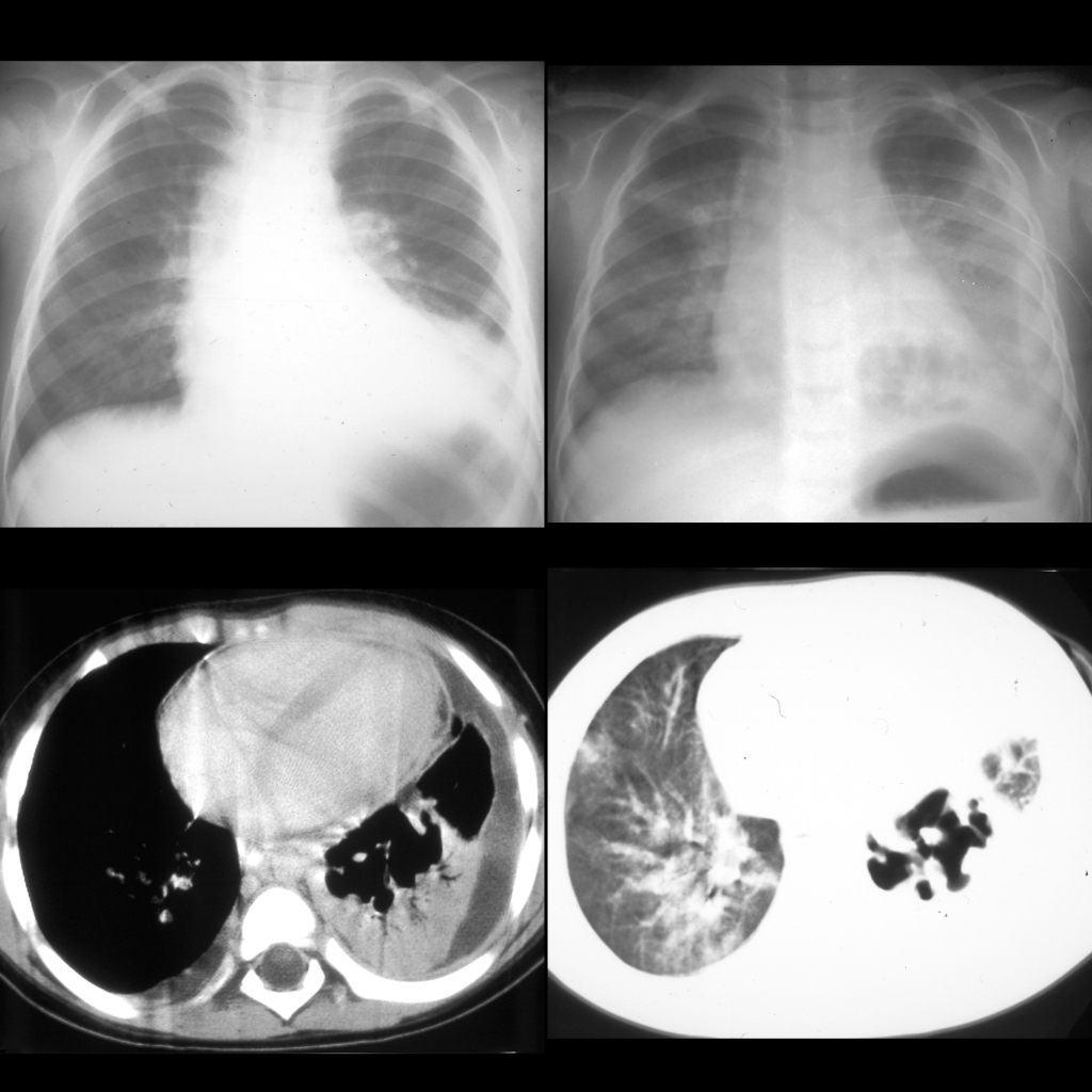
CXR AP (above left) initially shows an infiltrate in the left lower lobe that on a CXR AP one week later (above right) has developed a cystic cavity in the middle of it. A left pleural effusion is also now present. Axial CT with contrast of the chest (below) shows the cystic cavity is thin walled and septated. Left lower lobe consolidation and left pleural effusion remain present.
The diagnosis was post-infectious pneumatocele due to pneumococcal pneumonia.