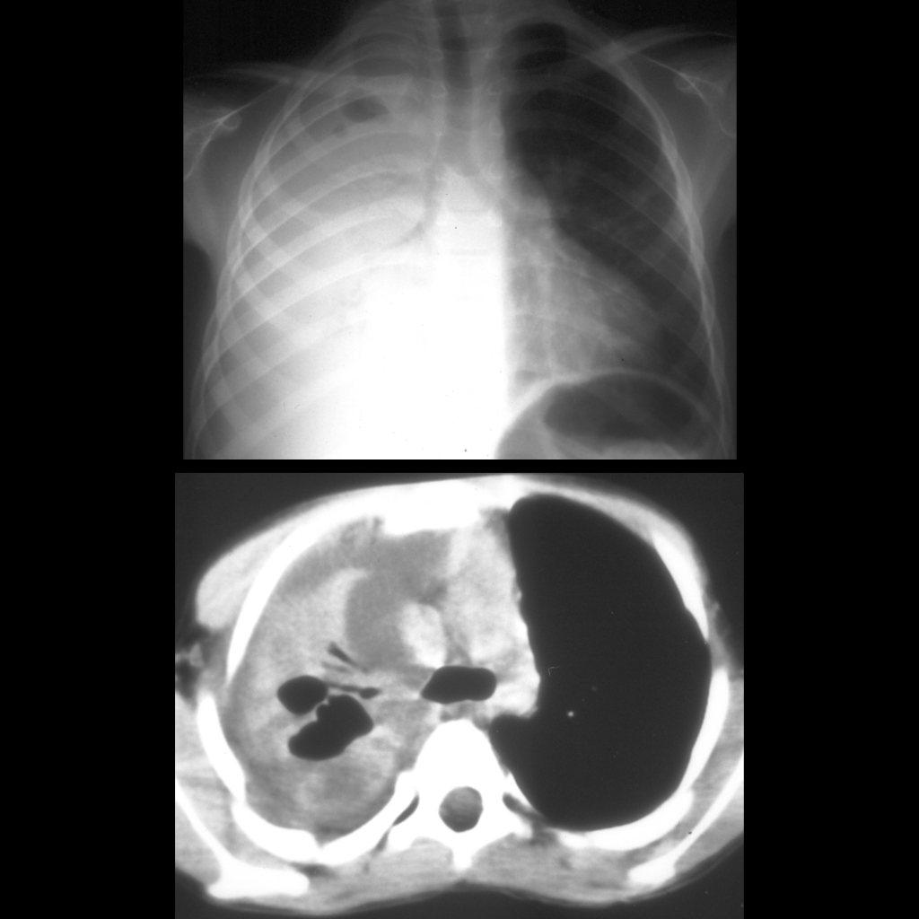
CXR AP (above) shows two air filled cavities in the right upper lobe in an otherwise completely opacified right hemithorax. Axial CT with contrast of the chest (below) shows a large right pleural effusion, right lung consolidation and a cavitary lesion in the right upper lobe.
The diagnosis was a developing lung abscess due to Streptococcal pneumonia.