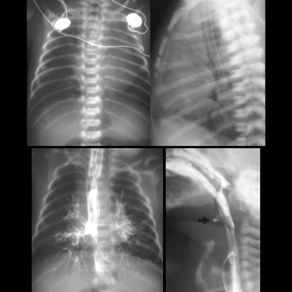
CXR AP and lateral (above) shows a nasogastric tube with its tip in the mid-esophagus. AP image from an UGI exam performed by injecting the nasogastic tube (below left) shows simultaneous opacification of the tracheobroncial tree and esophagus. Lateral image from the UGI exam (below right) shows the atretic origin of the trachea (black arrow). The tip of the endotracheal tube (which is in the esophagus) is at approximately the same level.