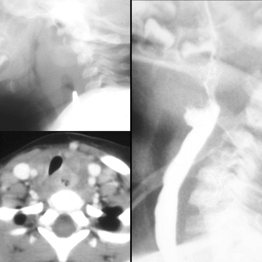
Lateral radiograph of the airway (above left) shows a radioopaque coin on edge in the esophagus at the level of C5. There is retropharygeal soft tissue swelling around the foreign body and air anterior to the coin in the retropharyngeal soft tissue. Axial CT with contrast of the neck after foreign body removal (below left) better shows the retropharyngeal fluid and air collection anterior to the vertebral body and causing some mass effect on the trachea. Lateral image from an upper GI exam (right) shows that the esophagus communicates with the retropharyngeal fluid and air collection.