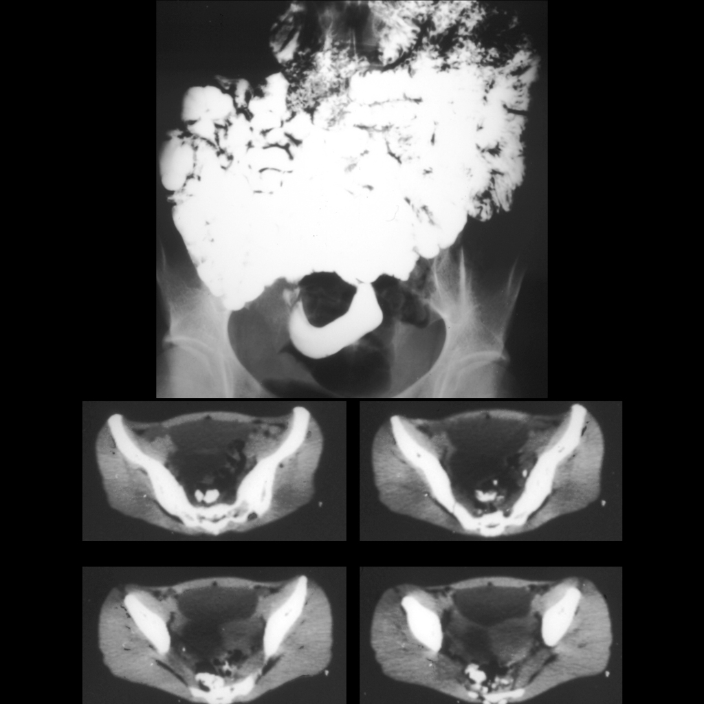
Image from a small bowel follow through exam (above) shows the small bowel to be entirely opacified and to be elevated out of the pelvis with the exception of a single loop in the middle of the pelvis. Axial CT without intravenous and with oral contrast of the abdomen (below) shows a large low density multiseptated mass in the pelvis.