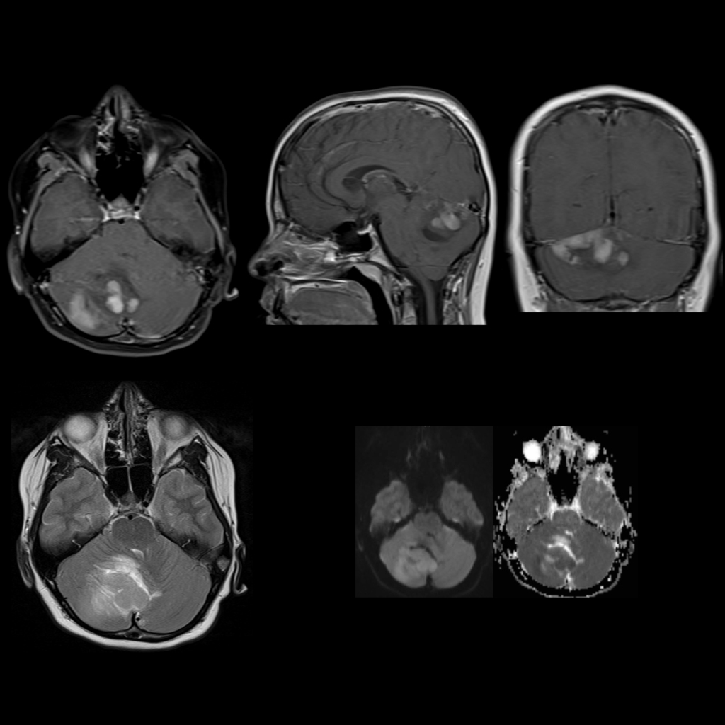
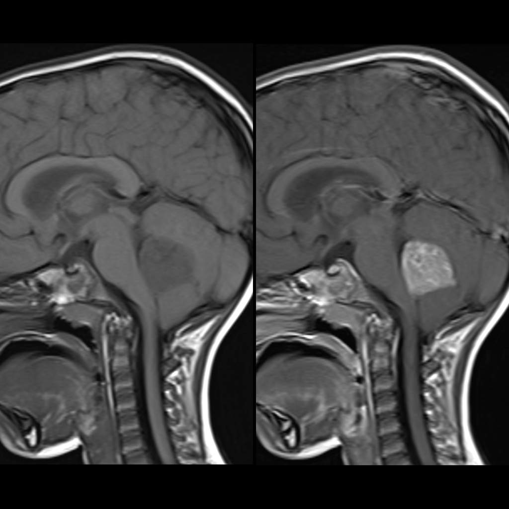
- Etiology: small round blue cell tumor
- Imaging: well defined / homogenous / enhancing mass, usually arising in midline in roof of 4th ventricle / inferior medullary velum, , can originate off midline such as from Foramen of Lushka, hydrocephalus
- CT: well defined and dense
- MRI: T1+T2 isointense, homogenous enhancement in 90-95%, calcifications and cyst rare, often restricted diffusion due to high degree of cellularity / high nuclear to cytoplasmic ratio / scant interstitial fluid / decreased extra-cellular space / can be useful for followup / monitoring
- DDX:
- Complications: systemic metastases in 15%, subarachnoid metastases in 50% – indistinct folia / sulci on T1, leptomeningeal enhancement, sugar coating of cord / drop metastases
- Treatment:
- Clinical: peaks in childhood and early adult
Radiology Cases of Medulloblastoma


