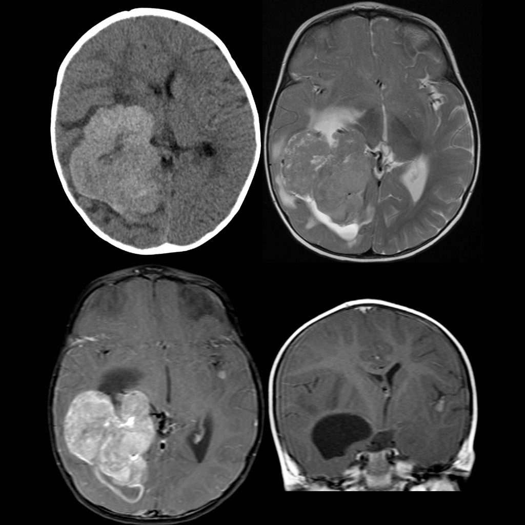
Axial CT without contrast of the brain (above left) shows a large high density mass that contains a few punctate calcifications in the trigone of the right lateral ventricle that is causing midline shift to the left. Axial T2 MRI (above left) of the brain shows the mass to surrounded by edema and to be causing some transependymal flow of cerebrospinal fluid. Axial (below left) and coronal (below right) T1 MRI with contrast show homogenous enhancement of the mass. A separate enhancing nodule is also noted in the left temporal lobe in the region of the left Sylvian fissure.