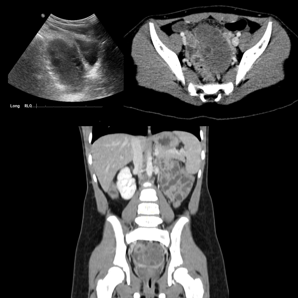
Sagittal US of the pelvis (above left) shows an enlarged right ovary with multiple peripheral follicles. Axial CT with contrast of the abdomen (above right) shows the right ovary to be enlarged with multiple peripheral follicles and to be malpositioned in the midline of the pelvis while the coronal CT (below) shows the right ovary to be in a position in the midline above the bladder.