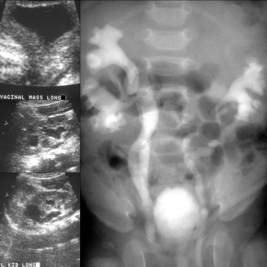
Newborn with oligohydramnios and hydroureteronephrosis on prenatal US

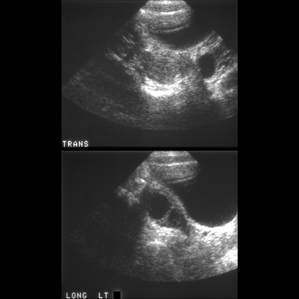
US of cystic renal dysplasia
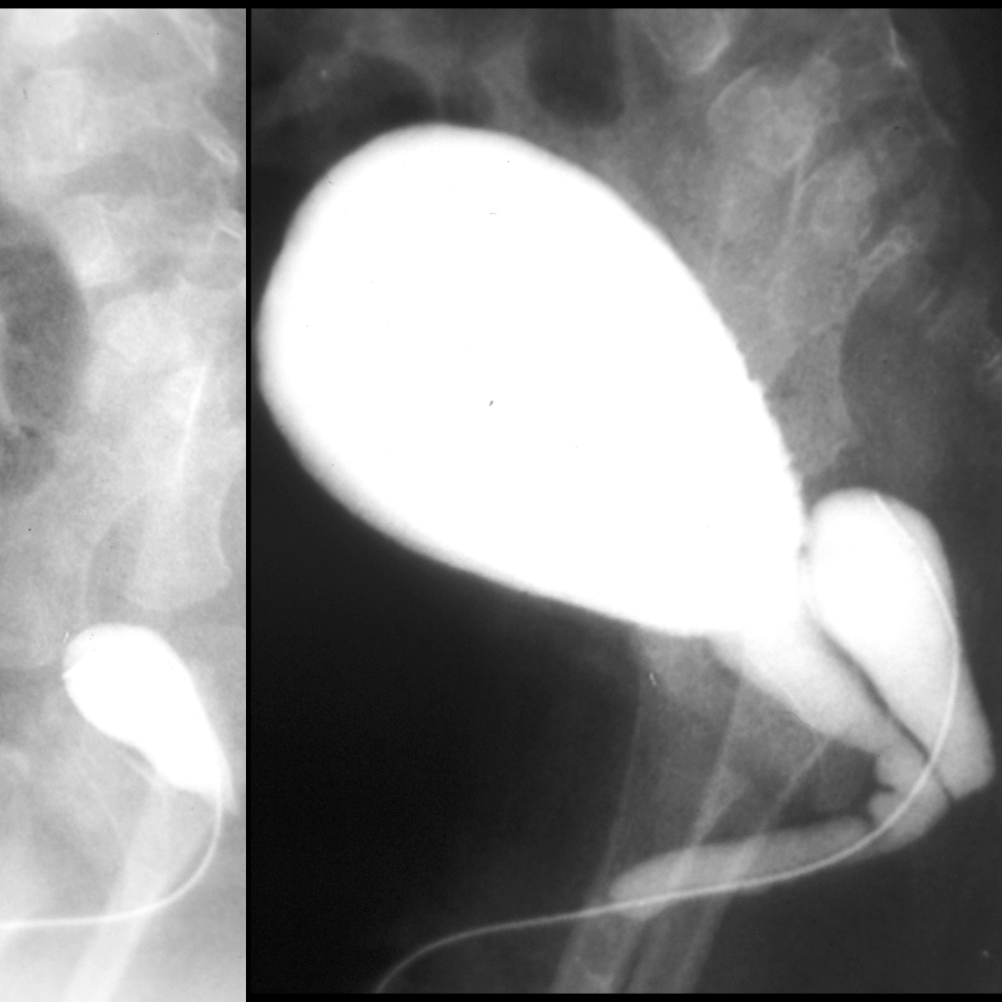
Sagittal US of the kidneys (above) show small echogenic kidneys bilaterally with some cysts but no hydronephrosis. AP image from a VCUG (below left) shows bilateral grade 4 vesicoureteral reflux with intrarenal reflux and the bladder is trabeculated. Lateral image from the VCUG (below right) shows a urachal diverticulum arising from the dome of the bladder anteriorly. The urethra was normal.

The diagnosis was vesicoureteral reflux causing cystic renal dysplasia and an incidental urachal remnant.

School ager with hypertension and neurofibromatosis Type 1

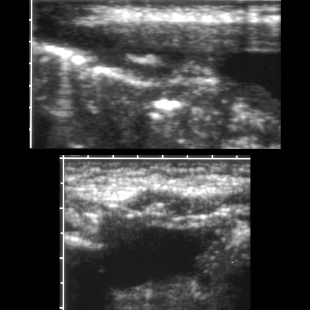
CT angiogram and spectral doppler US of bilateral renal artery stenosis in a patient with neurofibromatosis Type 1
3D reconstruction of a CT angiogram with contrast of the chest and abdomen (left) shows diffuse moderate narrowing of the mid aorta and marked narrowing of the origins of the renal arteries bilaterally. Spectral doppler US of the renal arteries (right) shows parvus tardis waveforms within them bilaterally.

The diagnosis was bilateral renal artery stenosis in a patient with neurofibromatosis Type 1.

Newborn with a left abdominal mass and palpable subcutaneous nodules on the right chest wall, left thigh, and right buttock

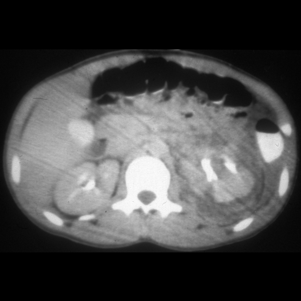
CT of neuroblastoma Stage 4S
Coronal (above left), sagittal (above right) and axial (below left) CT with contrast of the abdomen show a large left-sided suprarenal mass which is calcified and displaces the right kidney inferiorly and posterior. Axial CT of the pelvis (below right) shows the small right gluteal subcutaneous soft tissue nodule.

The diagnosis was neuroblastoma Stage 4S.

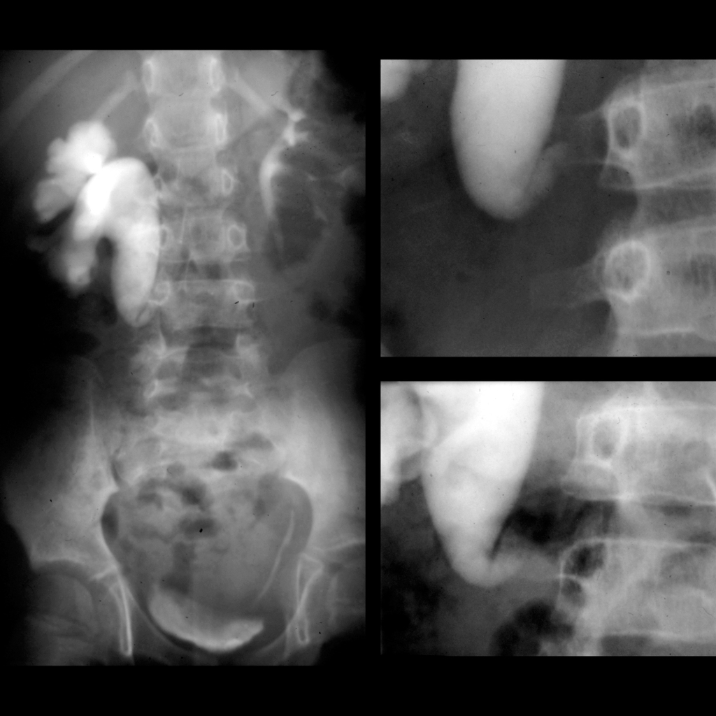
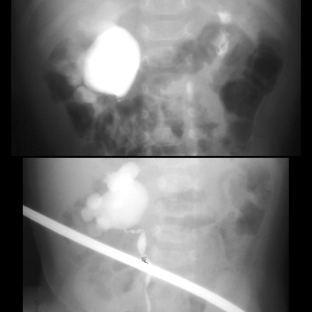
Teenager with pelvic fracture from trauma who cannot void

VCUG and RUG of urethral transection
AP (upper left) and oblique (upper right) images from a voiding cystourethrogram with the bladder filled through a suprapubic catheter shows extravasation of contrast into the base of the penis when the patient attempts to void. Oblique image from a retrograde urethrogram (below) shows an inability to fill the urethra completely during retrograde injection.

The diagnosis was urethral transection confirmed at cystoscopy.

Teenager with enuresis

US of urethral trauma
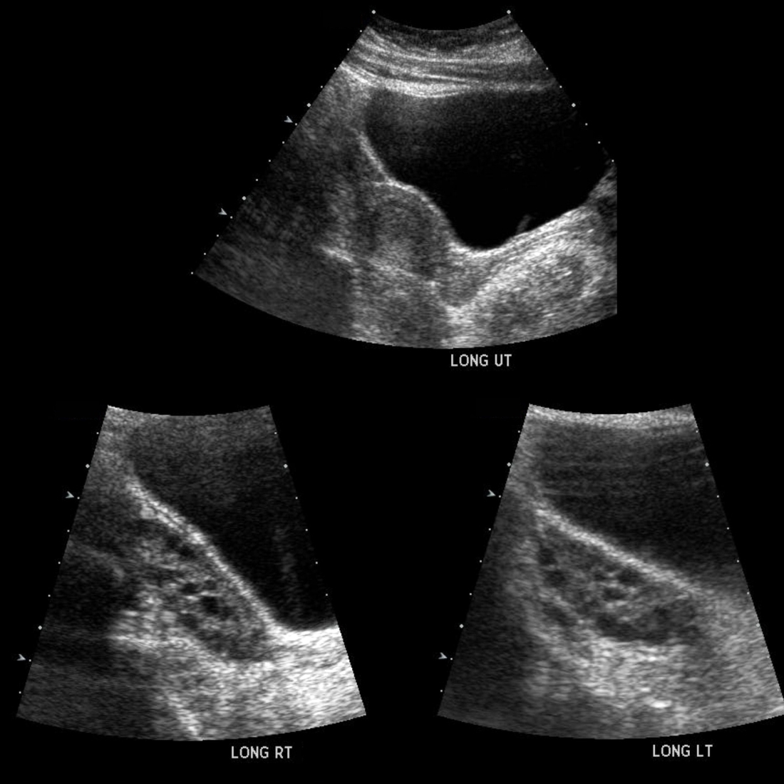
Transverse US of the prostatic urethra (upper left) shows a small round echogenic lesion with posterior shadowing in the center of the prostatic urethra which was not seen in the transverse US of the penile urethra (upper right). Sagittal US of the bladder (lower left) shows an echogenic lesion with posterior shadowing in the posterior aspect of the bladder that on transverse US of the bladder (lower right) resolved into two separate echogenic lesions with posterior shadowing.

The diagnosis was urethral trauma due to a metal axle from a toy car the patient had inserted into his urethra years before which had perforated the bladder creating a colovesical fistula and caused development of two bladder stones.

School ager with anti-NMDA encephalitis

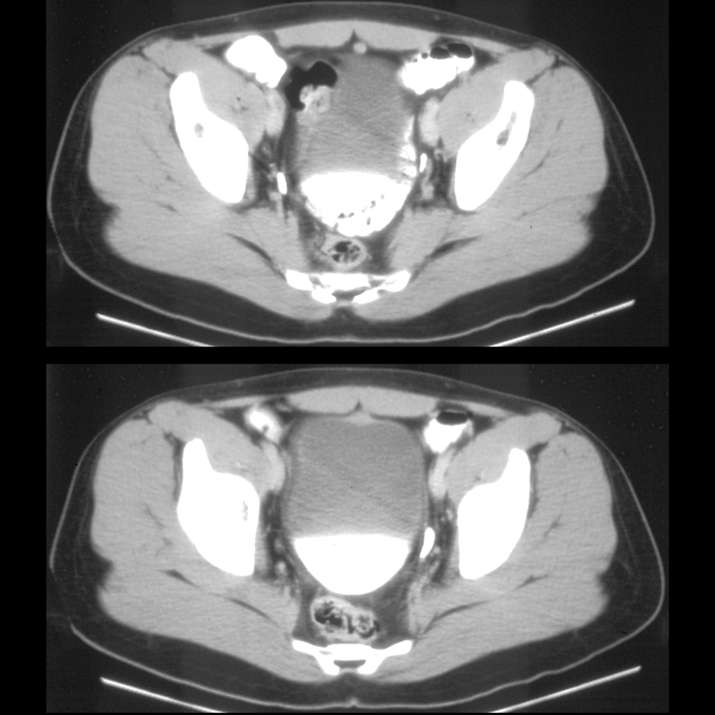
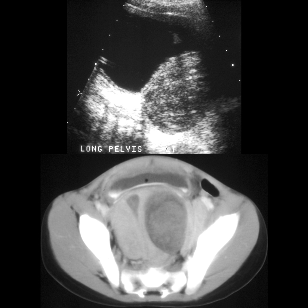
US and CT of ovarian teratoma in NMDA encephalitis
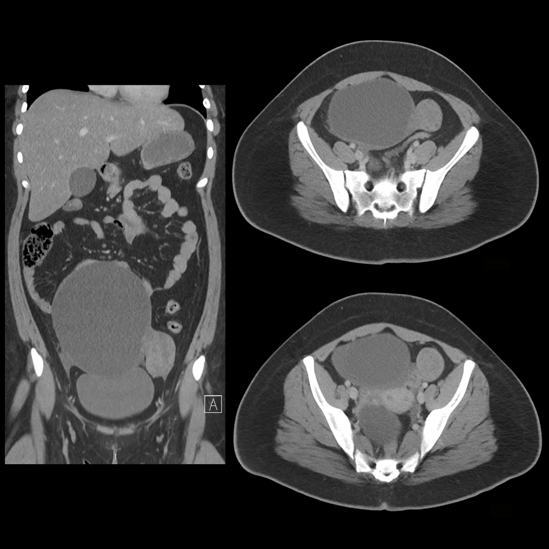
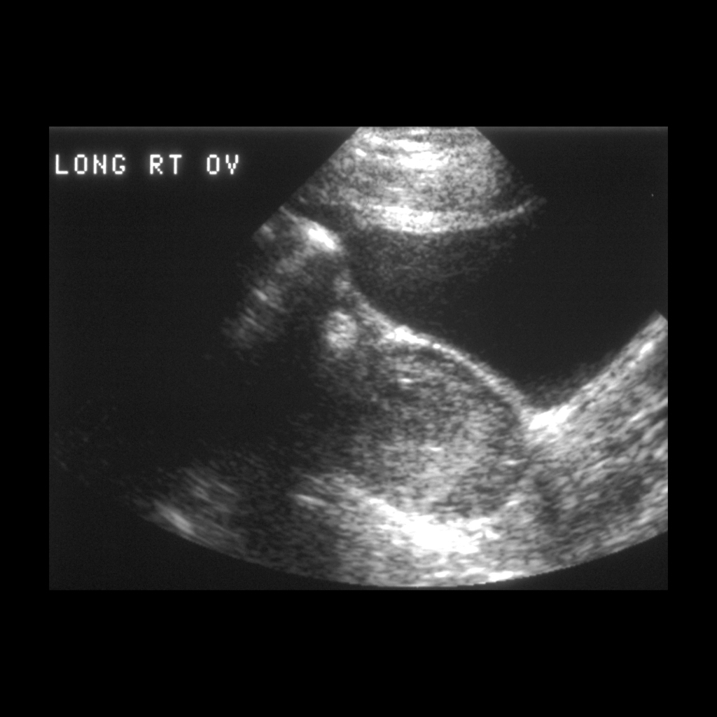
Sagittal US of the pelvis (upper left) shows the right ovary to be enlarged and to contain an echogenic lesion within it superiorly. Axial (upper right) and coronal (lower left) and sagittal (lower right) CT with contrast of the abdomen shows an enlarged right ovary in the midline of the pelvis anterior to the rectum which has multiple low density lesions within it.

The diagnosis was ovarian teratoma.