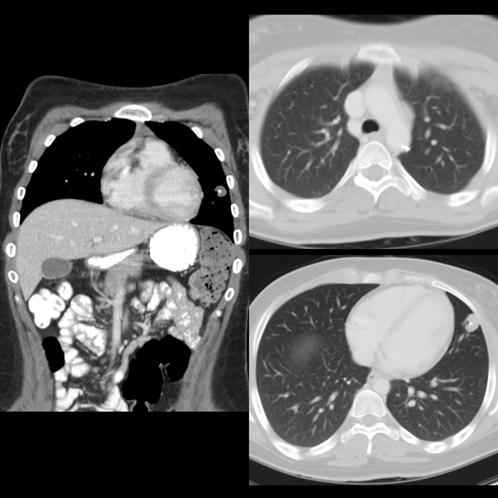
Coronal CT with contrast of the chest (left) shows a large round soft tissue nodule with a calcified center in the lateral aspect of the left hemithorax. Axial CT (above right) shows calcified mediastinal lymph nodes near the descending aorta and better demonstrates the calcification in the lung nodule (below right).