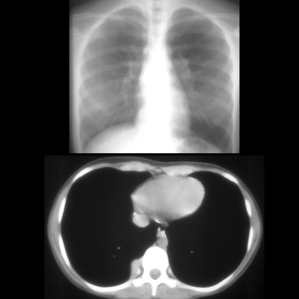
Teenager with past history of Wilms tumor CXR AP (above) shows a soft tissue density projecting in the right cardiophrenic angle. Axial CT with contrast of the chest (below) shows a soft tissue mass in the right posterior costophrenic sulcus. The diagnosis was lung metastasis in a patient with Wilms tumor.