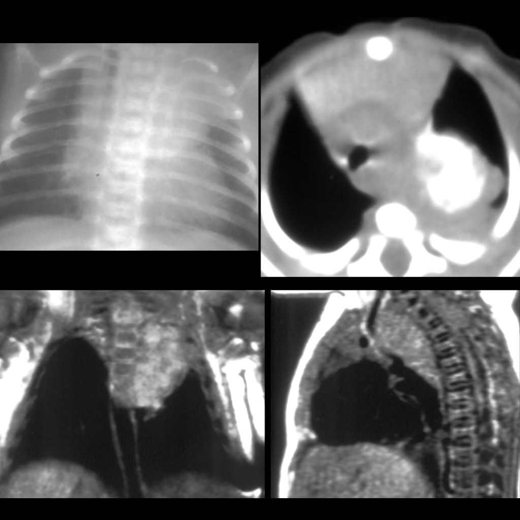
CXR AP (above left) shows a soft tissue mass in the left superior chest causing spreading and erosion of the left first and second rib, and increased distance between the left C7 and T1 transverse processes. Axial CT without contrast of the chest (above right) shows the mass to be densely calcified, in the posterior mediastinum, and displacing and compressing the trachea. Coronal (below left) and sagittal (below right) T1 MRI without contrast of the chest show a posterior mediastinal mass from C7 to T3 that has intraspinal extension.