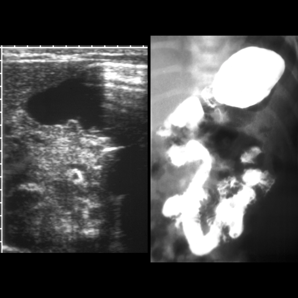
Transverse US of the pylorus (left) showed a normal appearing pylorus and showed the superior mesenteric vein to be directly above the superior mesenteric artery (the round structure with an echogenic rim in the center of the image), raising suspicion for malrotation. AP image from an UGI exam (right) shows the duodenal jejunal junction to be over the right pedicle of the spine and to be below the level of the duodenal bulb.