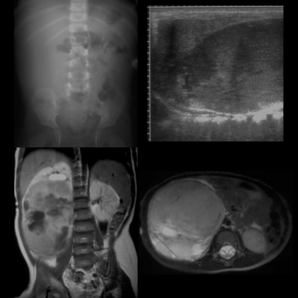
AXR (above left) shows displacement of the bowel out of the right side of the abdomen. Sagittal US of the right kidney (above right) shows a large right renal mass that spares the upper pole of the right kidney. Coronal T1 MRI with contrast of the abdomen (below left) shows a large mass that is heterogenous in appearance that arises from the lower pole of the right kidney and that is demonstrating a claw sign superiorly. Axial T2 MRI (below right) again shows the heterogenous nature of the mass due to hemorrhage and necrosis.