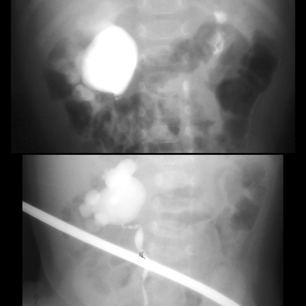
AP image from the excretory phase of an intravenous pyelogram (above) shows a normal left renal collecting system and a markedly dilated right renal collecting system. No contrast was seen in the right ureter. AP image from a retrograde pyelogram (below) shows a markedly dilated right renal collecting system and a narrowing and tortuosity to the proximal right ureter.