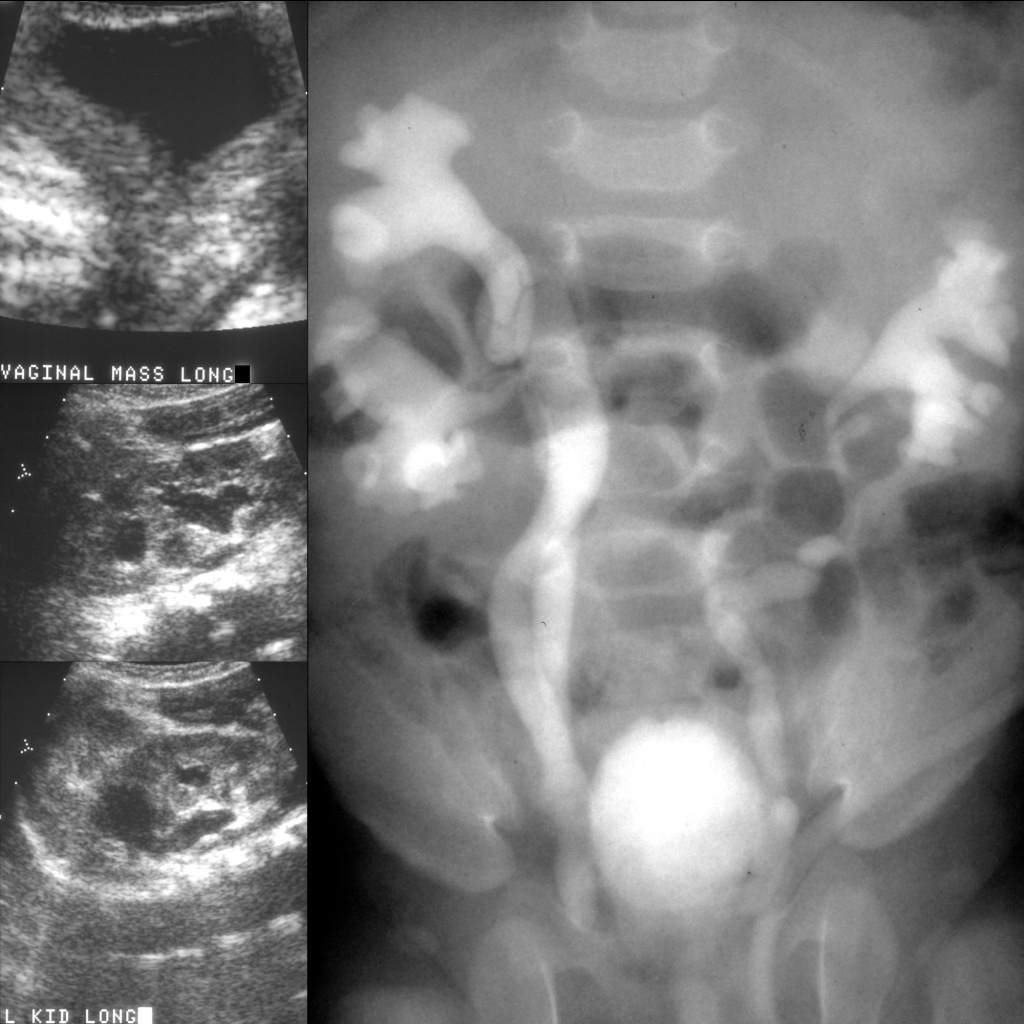
Sagittal US of the vaginal mass (above left) shows it to be cystic in nature. Sagittal US of the right kidney (middle left) shows moderate hydronephrosis of the upper and lower poles of a duplicated renal collecting system. Sagittal US of the left kidney (below left) shows marked hydronephrosis of the upper pole and moderate hydronephrosis of the lower pole of a duplicated renal collecting system. Excretory phase of a vintage intravenous pyelogram (right) shows on the right a moderately hydronephrotic duplicated renal collecting system and on the left an obstructed nonopacified nonfunctional upper pole collecting system which displaces the opacified functional moderately hydronephrotic lower pole collecting system inferolaterally (drooping lily sign).