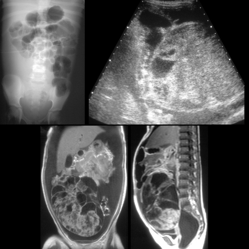
AXR (above left) shows displacement of the bowel loops to the left upper quadrant. Sagittal US of the abdomen (above right) shows a large solid heterogenous intraperitoneal mass in the lower right abdomen whose organ of origin was uncertain. There was a large amount of ascites. Coronal (below left) and sagittal (below right) T1 MRI with contrast of the abdomen shows a large right-sided mass that was oval in shape with smooth contour with multiple cystic lobulations with enhancing septae within it superiorly with it being more solid inferiorly and which appeared to be adherent to the right ovary.