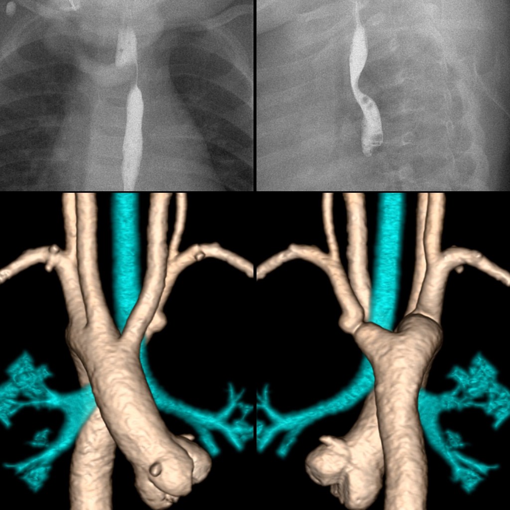
AP image from an upper GI exam (above left) shows a right sided aortic arch and an indentation running at an angle from right to left across the upper esophagus. Lateral image from an upper GI exam (above right) shows a posterior indentation across the upper esophagus. 3D CT with contrast of the chest viewed from the front (below left) shows a right sided aortic arch and a right sided descending aorta and viewed from the back (below right) shows the aberrant left subclavian artery arising from a large diverticulum of Kommerell. The airway (in blue) was not compressed.