Teenager with black and blue subcutaneous nodules and chronic GI bleeding

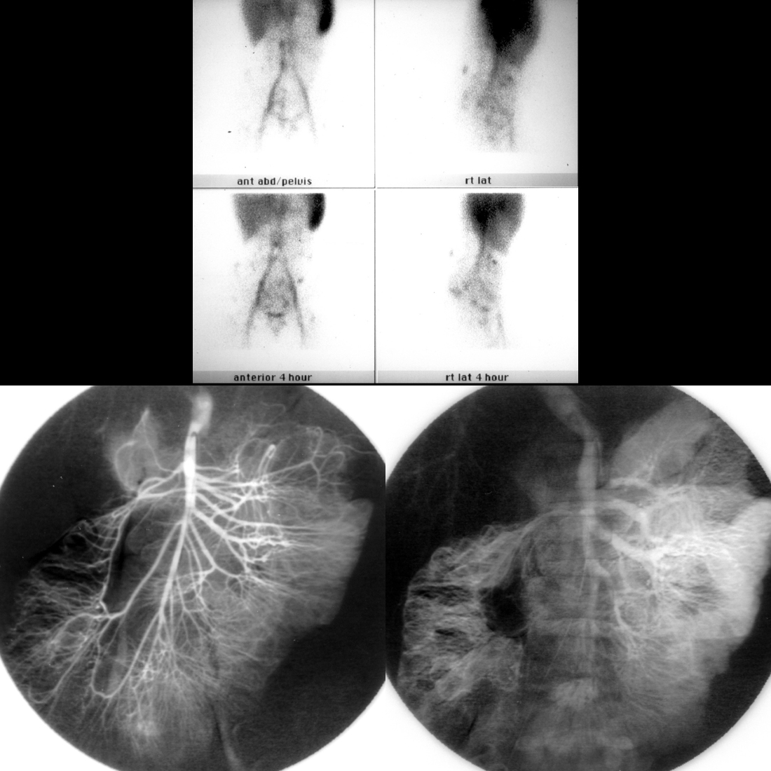
Tagged red blood cell nuclear medicine scan and angiogram of blue rubber bleb nevus syndrome
Nuclear medicine tagged red blood cell scan for GI bleeding shows AP and lateral images of the abdomen obtained immediately after the beginning of the exam (above top row) and 4 hours later (above bottom row). The images show immediate pooling of radiotracer in multiple discrete areas in the abdomen and pelvis with continued radiotracer accumulation in these areas over time. Arterial phase image from a superior mesenteric artery angiogram (below left) shows normal arterial vessels, while the venous phase (below right) shows contrast puddling in rounded spaces that filled in over time in the small bowel and ascending colon that were consistent with venous malformations.

The diagnosis was blue rubber bleb nevus syndrome.
Premature newborn with new left femoral vein catheter that will not infuse

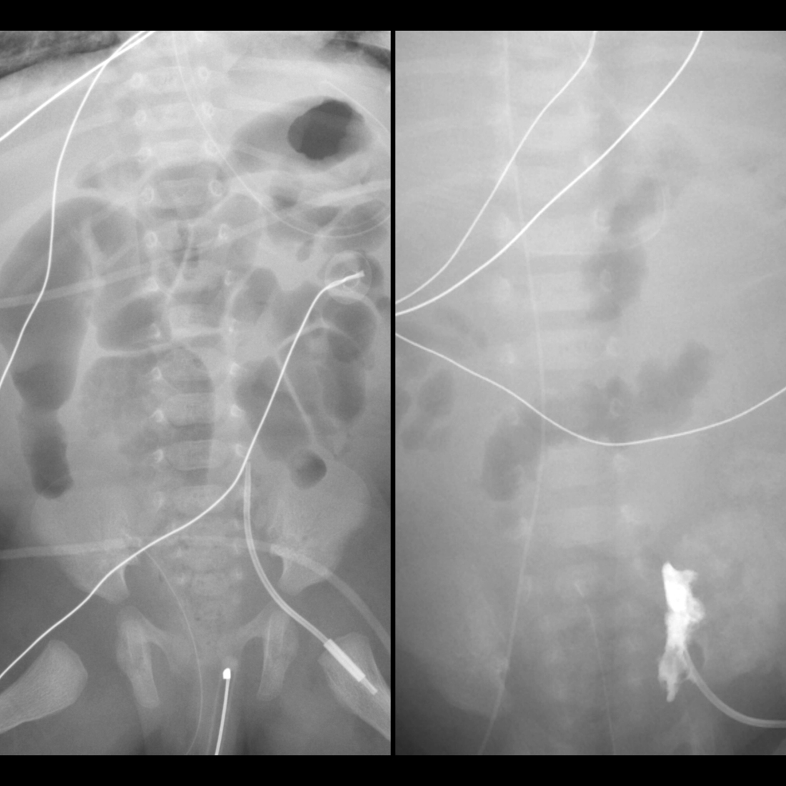
Venogram of perforation of femoral venous catheter out of the femoral vein
AXR AP (left) shows a left femoral venous catheter whose tip projects to the left of the L5 vertebral body, rather than projecting near the middle of the L5 vertebral body. AP venogram taken after injecting contrast through the femoral catheter (right) shows extravasation of contrast into the soft tissues surrounding the catheter tip.

The diagnosis was femoral venous catheter malfunction with perforation of the femoral venous catheter out of the femoral vein.
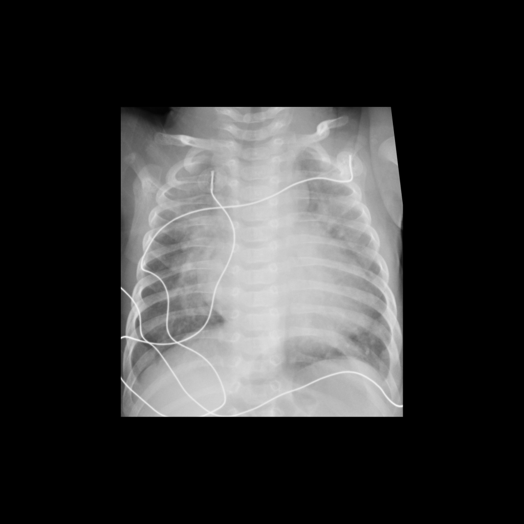
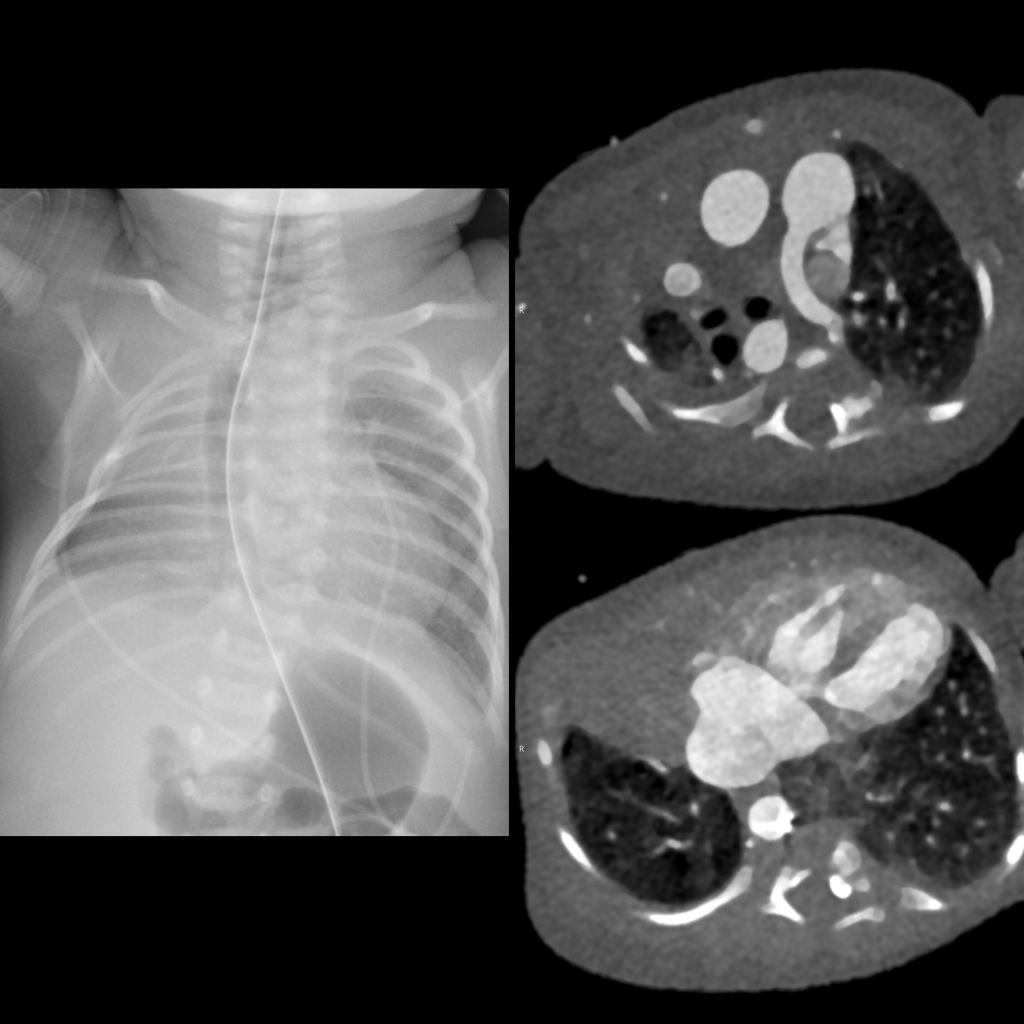
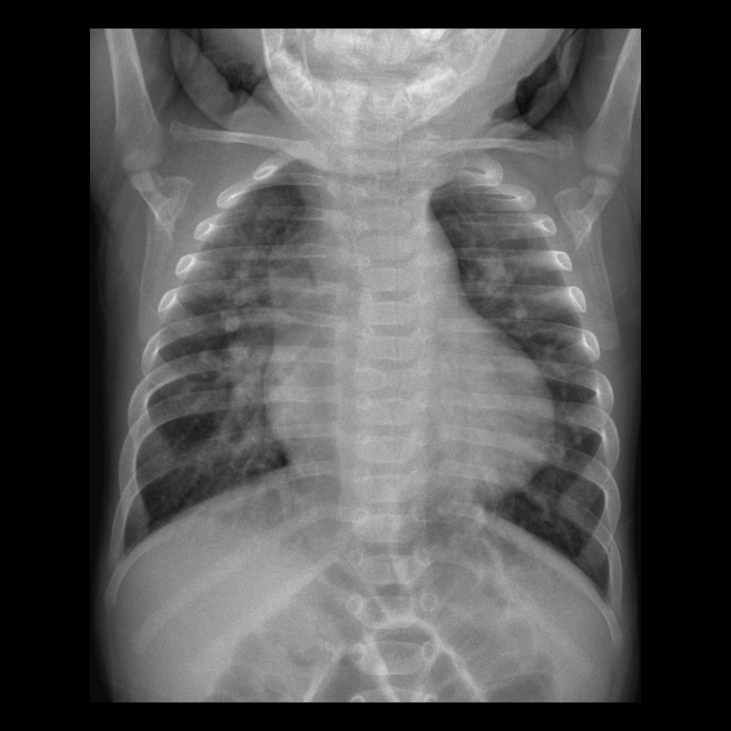
Newborn with respiratory distress

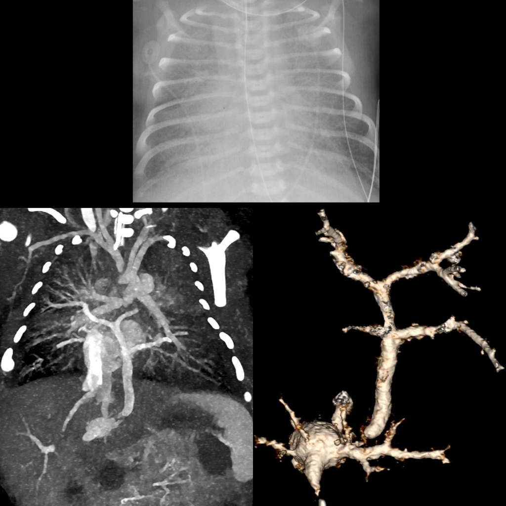
CXR and CT of infracardiac total anomalous pulmonary venous return
CXR AP (above) shows mild cardiomegaly with marked pulmonary vascular congestion. Coronal CT MIP with contrast of the chest (below left) shows the pulmonary veins from both lungs draining into the vertical vein which crosses the diaphragm and then narrows as it connects to the dilated ductus venosus which then drains into the inferior vena cava. This is better demonstrated on the 3D CT (below right).

The diagnosis was infracardiac total anomalous pulmonary venous return.
Infant with dysphagia

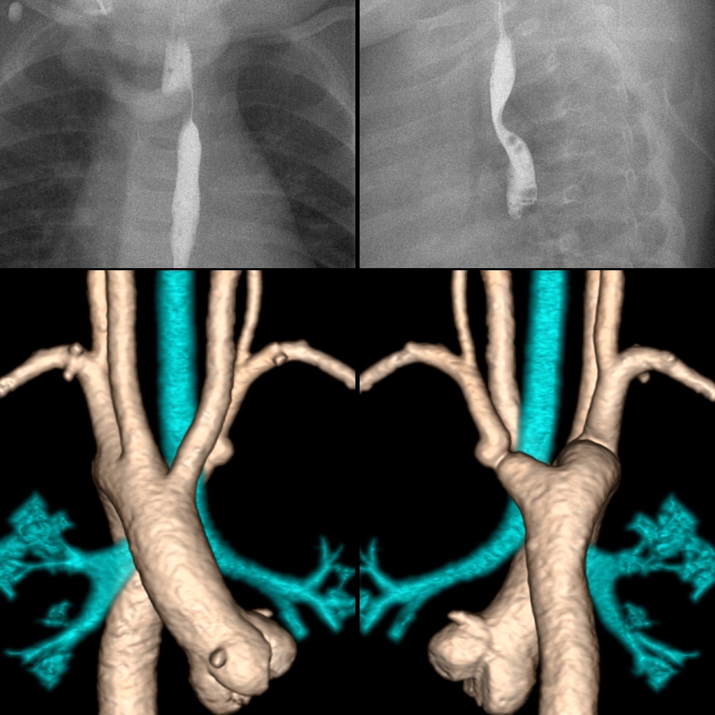
CT of right aortic arch with aberrant left subclavian artery
AP image from an upper GI exam (above left) shows a right sided aortic arch and an indentation running at an angle from right to left across the upper esophagus. Lateral image from an upper GI exam (above right) shows a posterior indentation across the upper esophagus. 3D CT with contrast of the chest viewed from the front (below left) shows a right sided aortic arch and a right sided descending aorta and viewed from the back (below right) shows the aberrant left subclavian artery arising from a large diverticulum of Kommerell. The airway (in blue) was not compressed.

The diagnosis was right aortic arch with aberrant left subclavian artery.

Newborn with complex congenital heart disease on prenatal US

CXR and CT of pulmonary agenesis
CXR AP (left) shows the right hemithorax to be smaller than the left hemithorax and there is opacity in the right upper and middle lobes and an aerated right lower lobe. There are multiple hemivertebrae and a butterfly vertebra in the thoracic spine causing a scoliosis convex left. Axial CT with contrast of the chest (above right) shows the right pulmonary artery to be absent along with the right upper lobe. Axial CT (below right) shows absence of the right middle lobe of the lung but the right lower lobe of the lung is present and aerated via a small bronchus off of the trachea (not shown) and perfused by collateral vessels arising from the 7 o’clock position off of the aorta.

The diagnosis was right pulmonary agenesis with the hypoplastic right lower lobe receiving its arterial supply from collaterals off of the aorta in a patient who has multiple vertebral body anomalies resulting in congenital scoliosis.

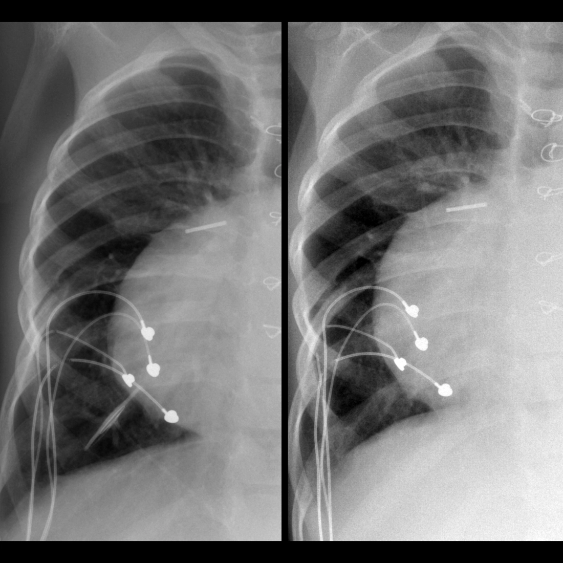
School ager with a central venous catheter that infuses but does not draw back

CXR of central venous catheter tip occlusion against a vessel wall
CXR AP from one month ago (above) shows the tip of the central venous catheter to be at the junction of the superior vena cava and the right atrium. CXR AP today (below) shows the tip of the central venous catheter to have migrated back into the right subclavian vein and the tip of the catheter is occluded against the wall of the subclavian vein.

The diagnosis was central venous catheter malfunction after the catheter tip migrated from the superior vena cava to the right subclavian vein and the tip became occluded against the wall of the right subclavian vein.

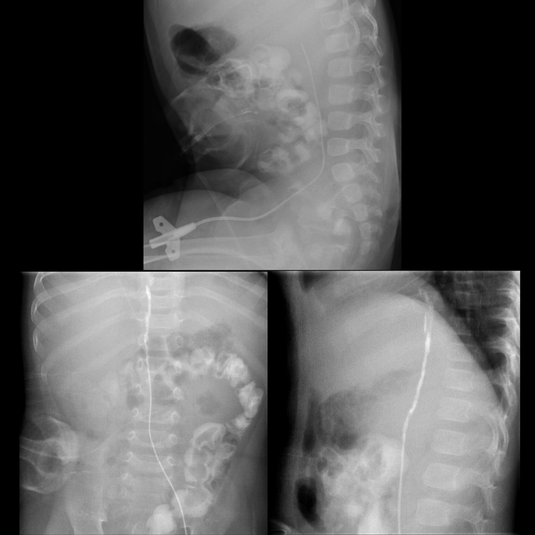
Newborn with respiratory distress on ECMO

CXR of malposition ECMO catheter in patient with congenital diaphragmatic hernia
CXR shows normal position of the arterial catheter tip in the aortic arch and abnormal position of the venous catheter tip (represented by a radio-opaque point) in the superior vena cava. Further advancement of the tip into the right atrium is necessary. Multiple cystic structures are present in the left hemithorax. The lungs are densely opacified.

The diagnosis was extracorporeal membrane oxygenation catheter malposition with the venous catheter tip in the superior vena cava in a patient with left sided congenital diaphragmatic hernia containing stomach and loops of bowel.

Preschooler with vomiting

UGI and CT of left aortic arch with aberrant right subclavian artery
AP image from an upper GI exam (above left) shows a left sided aortic arch and an indentation running at an angle from left to right across the upper esophagus. Lateral image from an upper GI exam (above right) shows a posterior indentation across the upper esophagus. Axial CT with contrast of the chest (below) shows an aberrant artery coursing anterior to the vertebral body and posterior to the aerated trachea and collapsed esophagus.

The diagnosis was left aortic arch with aberrant right subclavian artery.

Teenager after a motorcycle accident whose CXR showed a widened mediastinum

CT of thoracic aortic injury
Axial CT with contrast of the chest at the level of the aortic arch (above left) shows a large amount of fluid in the superior mediastinum while subsequent lower slices (above right and below left) show an intimal flap in the descending aorta. Sagittal CT (below right) shows a pseudoaneurysmal dilation of the aorta beginning at the aortic isthmus.

The diagnosis was thoracic aortic injury.

School ager with shortness of breath

CXR and CT of pericardial effusion due to histoplasmosis
CXR AP (above left) shows a large cardiac silhouette and an abnormal contour to the right superior mediastinum. Coronal CT with contrast of the chest (above right) shows a huge fluid collection in the pericardial space and a conglomeration of cystic lymph nodes in the right superior mediastinum. Axial CT (below) shows the pericardial fluid collection completely surrounding the heart and left lower lobe atelectasis.

The diagnosis was pericardial effusion due to histoplasmosis.

Preschooler with heartburn after eating

UGI and CT of left aortic arch with aberrant right subclavian artery
Lateral image from an UGI exam (above left) shows a persistent posterior indentation on the proximal esophagus. Sagittal CT with contrast of the chest (above right) shows an aberrant artery located posterior to the esophagus and anterior to a vertebral body. Axial CT (below) shows the right subclavian artery to be arising aberrantly from the aorta and coursing anterior to the vertebral body and posterior to the aerated trachea and collapsed esophagus.

The diagnosis was left aortic arch with aberrant right subclavian artery.

School ager with 1 week of abdominal pain and shortness of breath

CXR and US of pericardial effusion due to histoplasmosis
AXR (above left) shows hepatomegaly and an enlarged cardiac silhouette. CXR (above right) shows a water-bottle appearance to the cardiac silhouette and bilateral pleural effusions and bilateral hilar lymphadenopathy. Transverse US of the heart (below) shows a large anechoic fluid collection in the pericardial space.

The diagnosis was pericardial effusion due to histoplasmosis.