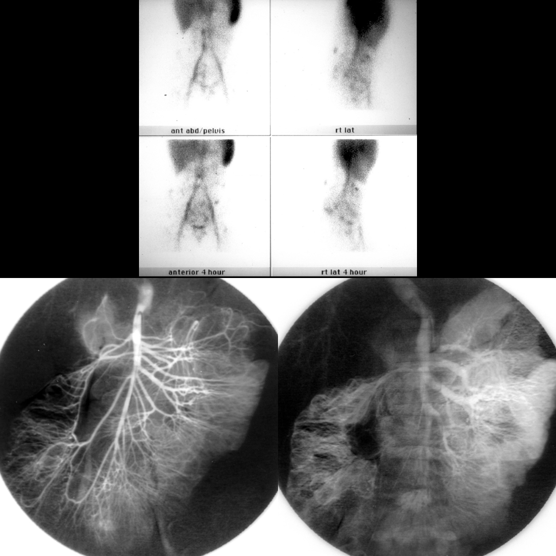
Nuclear medicine tagged red blood cell scan for GI bleeding shows AP and lateral images of the abdomen obtained immediately after the beginning of the exam (above top row) and 4 hours later (above bottom row). The images show immediate pooling of radiotracer in multiple discrete areas in the abdomen and pelvis with continued radiotracer accumulation in these areas over time. Arterial phase image from a superior mesenteric artery angiogram (below left) shows normal arterial vessels, while the venous phase (below right) shows contrast puddling in rounded spaces that filled in over time in the small bowel and ascending colon that were consistent with venous malformations.