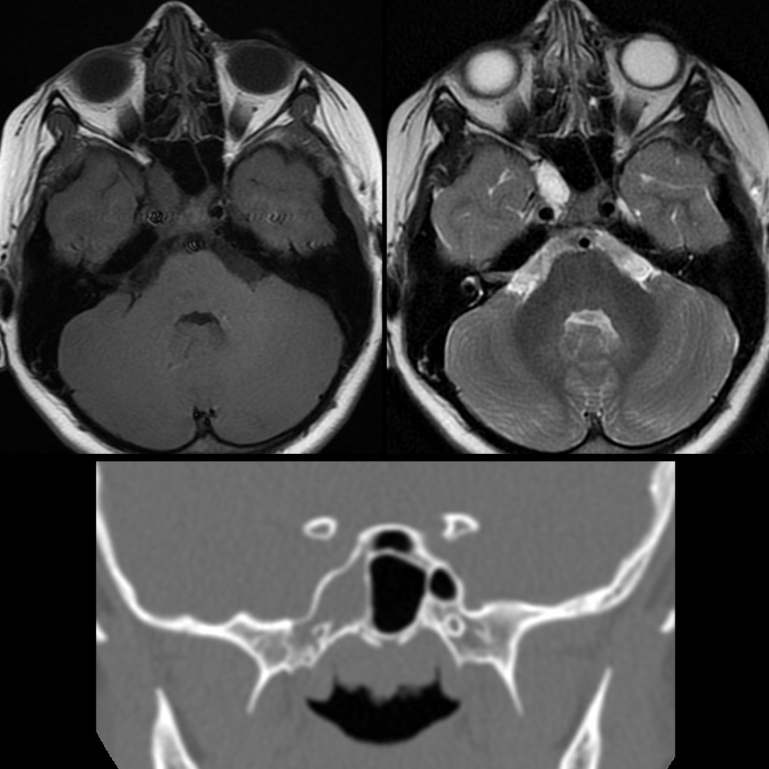
Axial T1 MRI without contrast of the brain (above left) shows an isointense signal intensity lesion in the right sphenoid bone that on T2 MRI (above right) shows a high signal intensity and which did not show enhancement post contrast. The remaining paranasal sinuses are clear. Coronal CT without contrast of the maxillofacial bones (below) shows the right sphenoid bone to be filled with a lucent non-expansile and non-destructive lesion.