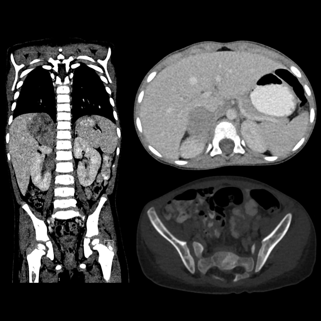
Coronal (left) and axial (above right) CT with contrast of the abdomen show a large low density faintly calcified mass arising from the right adrenal gland. Axial image of the pelvis in bone windows (below) shows multiple lytic bone lesions throughout the iliac wings and sacrum.