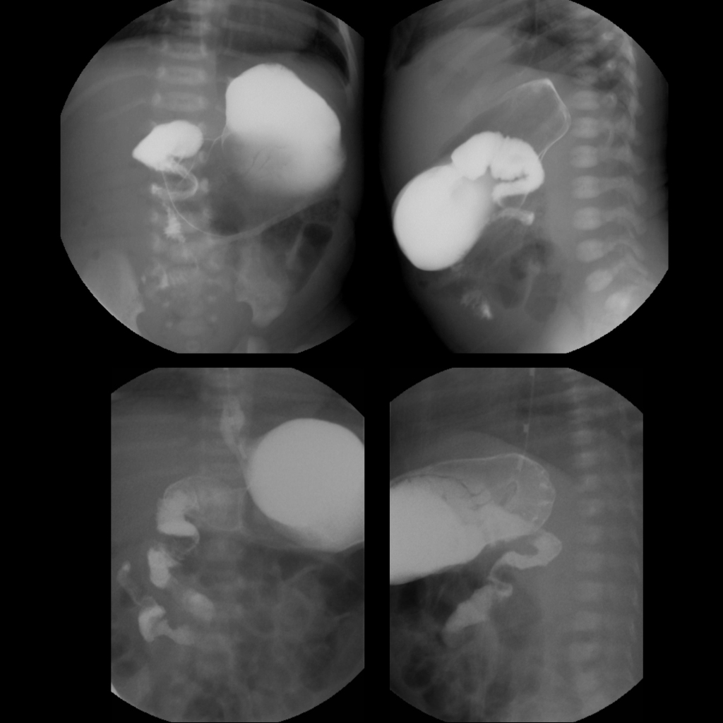
AP (above left) and lateral (above right) images from an upper GI exam on day of life 1 show dilation of the first part of the duodenum and a spiral or corkscrew appearance of the second and third parts of the duodenum with the ligament of Trietz projecting over the midline of the spine and lower than the first part of the duodenum. AP (below left) and lateral (below right) images from an upper GI obtained several days after a Ladd procedure show the first part of the duodenum now to be normal in caliber while the second and third parts of the duodenum continue to have a spiral or corkscrew appearance with the ligament of Trietz continuing to project over the midline of the spine and lower than the first part of the duodenum.