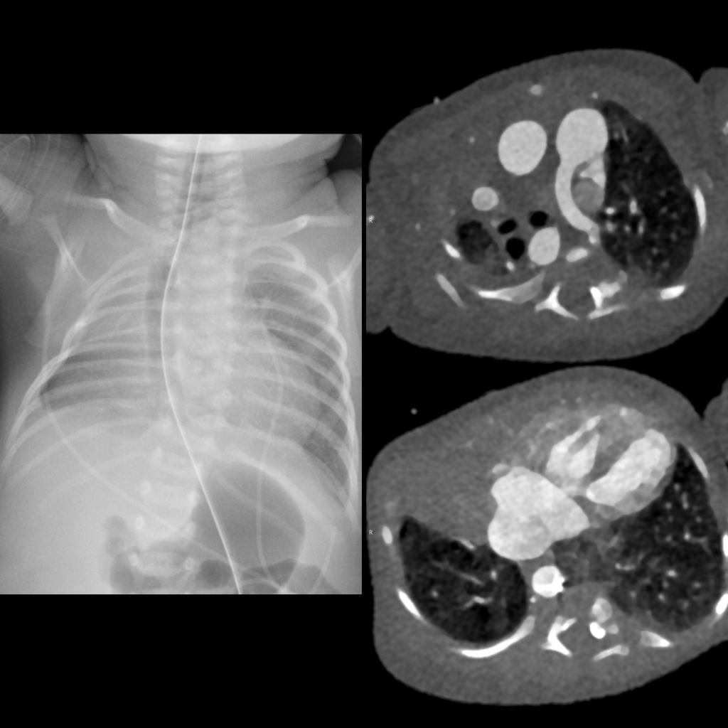
Newborn with complex congenital heart disease on prenatal US

CXR and CT of pulmonary agenesis
CXR AP (left) shows the right hemithorax to be smaller than the left hemithorax and there is opacity in the right upper and middle lobes and an aerated right lower lobe. There are multiple hemivertebrae and a butterfly vertebra in the thoracic spine causing a scoliosis convex left. Axial CT with contrast of the chest (above right) shows the right pulmonary artery to be absent along with the right upper lobe. Axial CT (below right) shows absence of the right middle lobe of the lung but the right lower lobe of the lung is present and aerated via a small bronchus off of the trachea (not shown) and perfused by collateral vessels arising from the 7 o’clock position off of the aorta.

The diagnosis was right pulmonary agenesis with the hypoplastic right lower lobe receiving its arterial supply from collaterals off of the aorta in a patient who has multiple vertebral body anomalies resulting in congenital scoliosis.