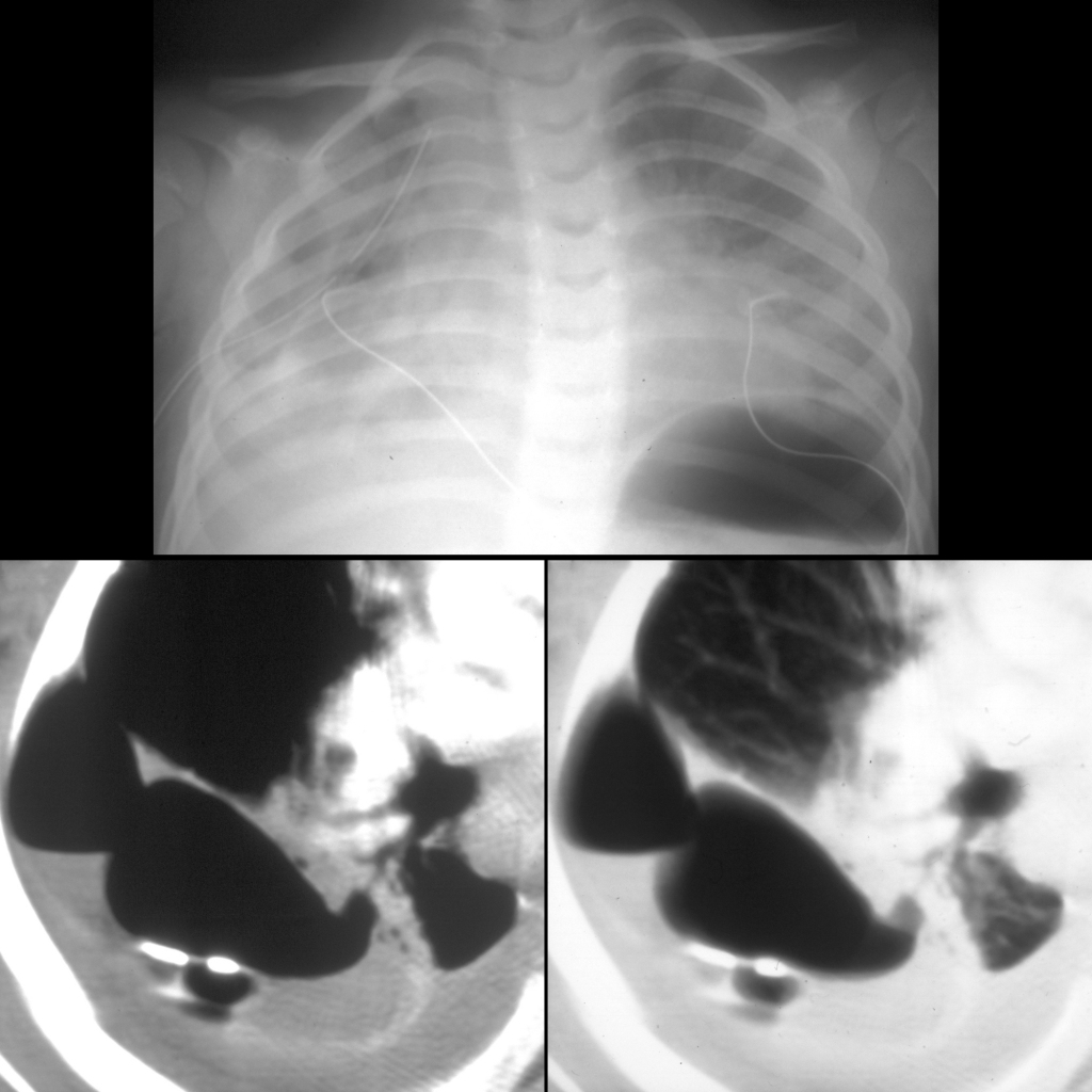
Toddler with respiratory distress

CXR and CT of post-infectious pneumatocele
CXR AP (above) shows the arterial catheter tip projecting over the aortic arch and the venous catheter tip projecting over the right atrium. There is bilateral diffuse airspace disease and bilateral pleural effusions. A cystic lesion is seen in the right lower lobe. Axial CT without contrast of the chest (below left) shows the cystic lesion is thin walled and communicates with the bronchial tree and has multiple septations (below right).

The diagnosis was pneumatocele due to pneumococcal pneumonia in a patient on ECMO.
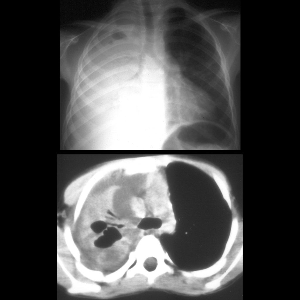
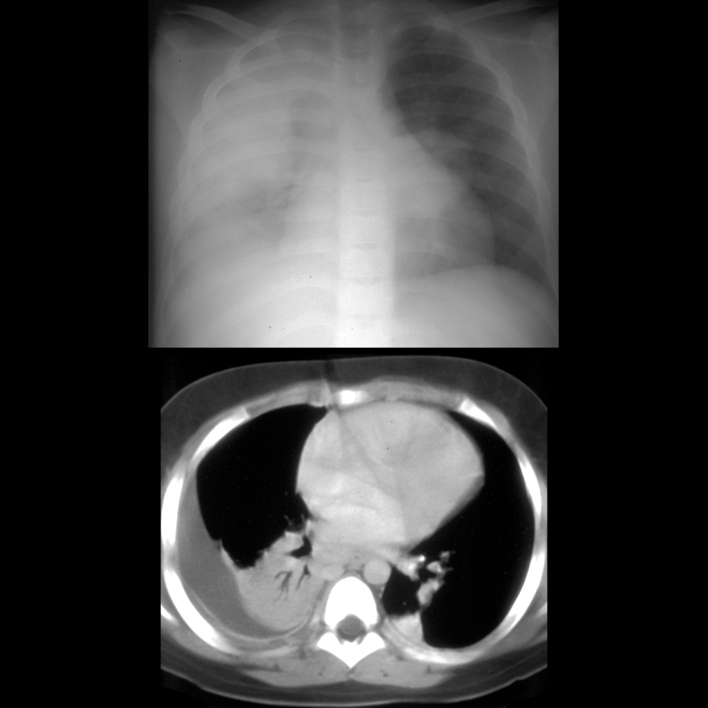
Toddler with respiratory distress

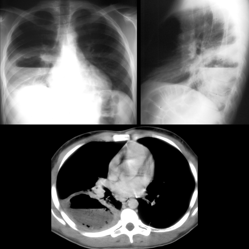
CXR and CT of bronchopleural fistula
CXR AP (above) shows diffuse bilateral airspace disease and a right sided pneumothorax that is almost completely drained by a right chest tube. Axial CT with contrast of the chest (below left) shows air and fluid and a chest tube in the right pleural space and an enhancing rim sign of the pleura. There is also a bronchopleural fistula from the superior segment of the right lower lobe to the necrotic lung and pleural space (below right).

The diagnosis was bronchopleural fistula and pleural empyema in a patient with streptococcus pneumonia.

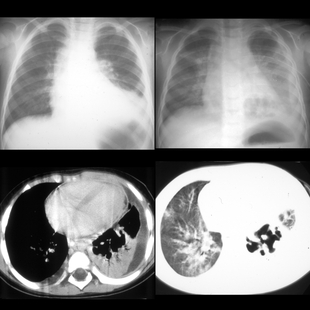
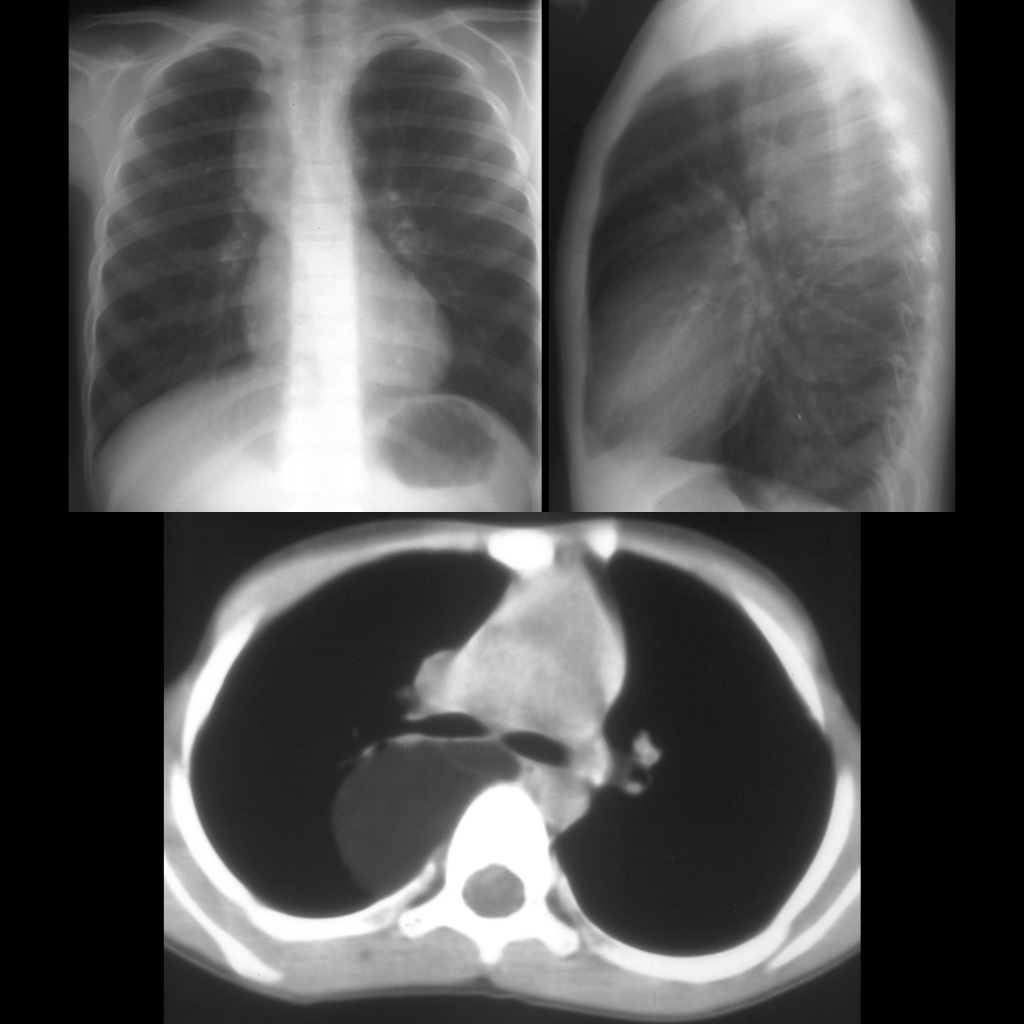
Toddler with respiratory distress

CXR and CT of post-infectious pneumatocele
CXR AP (above left) initially shows an infiltrate in the left lower lobe that on a CXR AP one week later (above right) has developed a cystic cavity in the middle of it. A left pleural effusion is also now present. Axial CT with contrast of the chest (below) shows the cystic cavity is thin walled and septated. Left lower lobe consolidation and left pleural effusion remain present.

The diagnosis was post-infectious pneumatocele due to pneumococcal pneumonia.
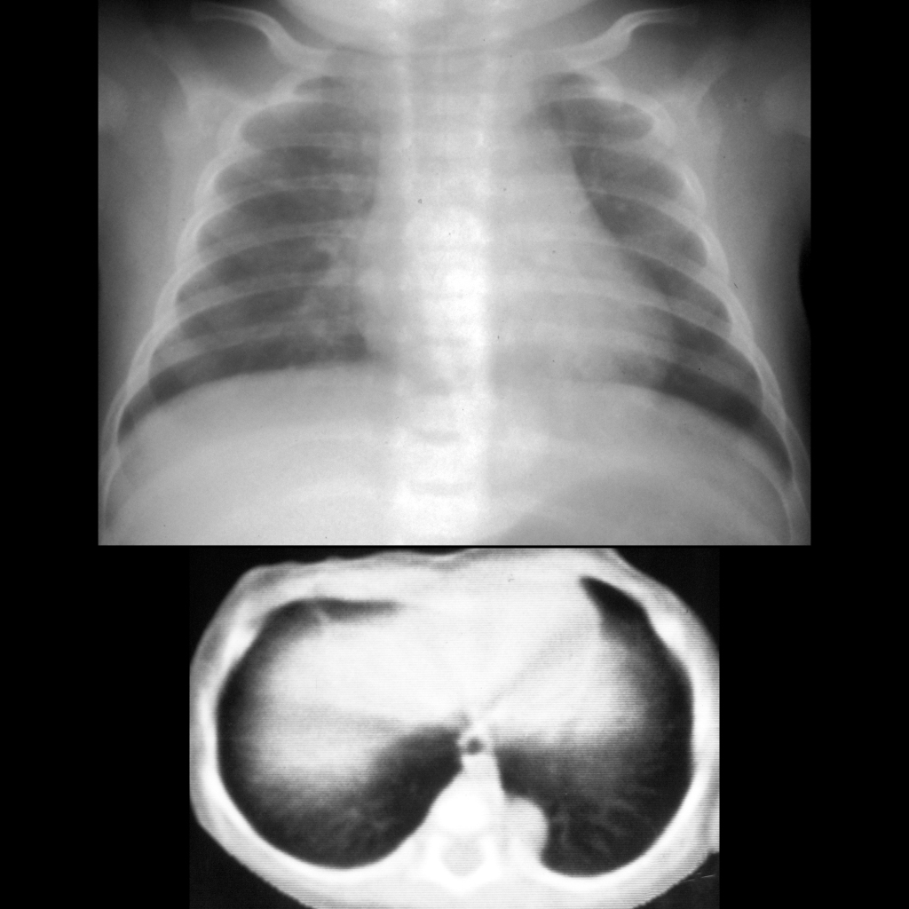
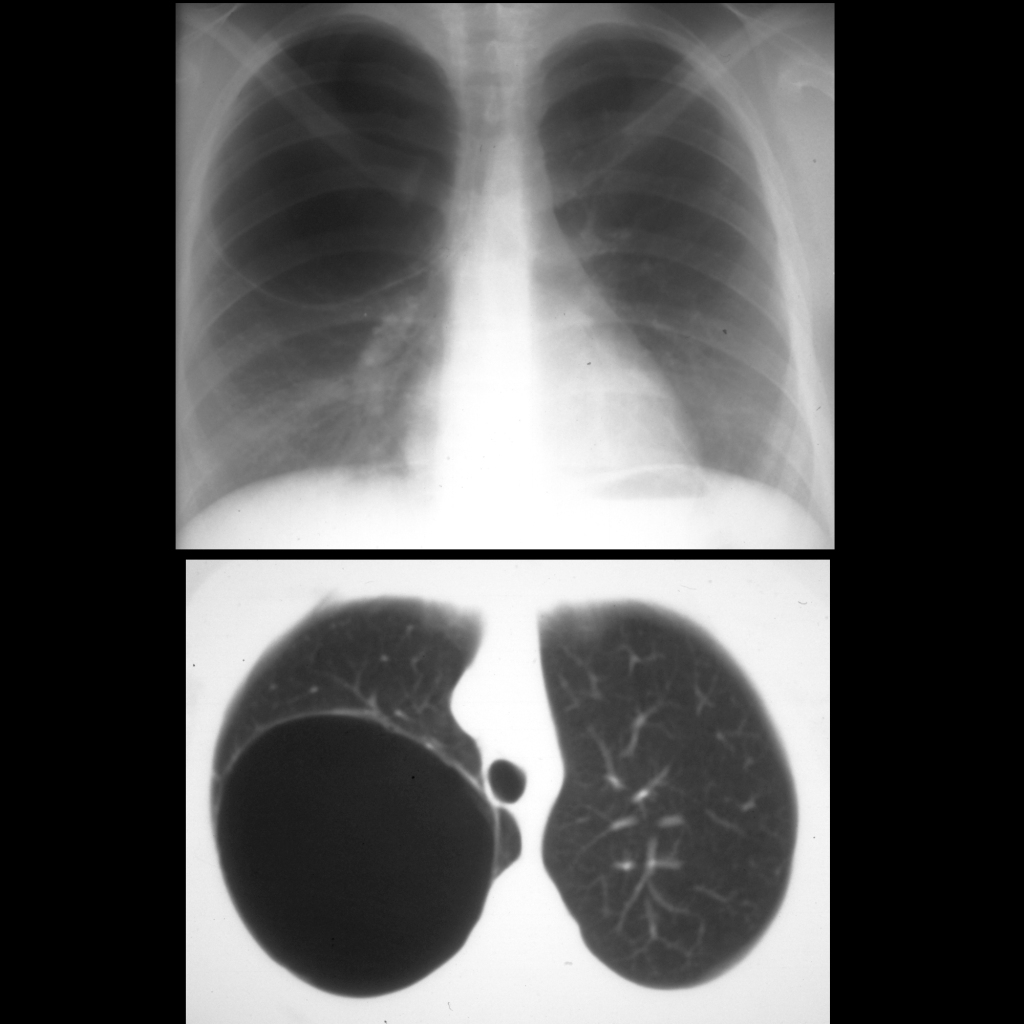
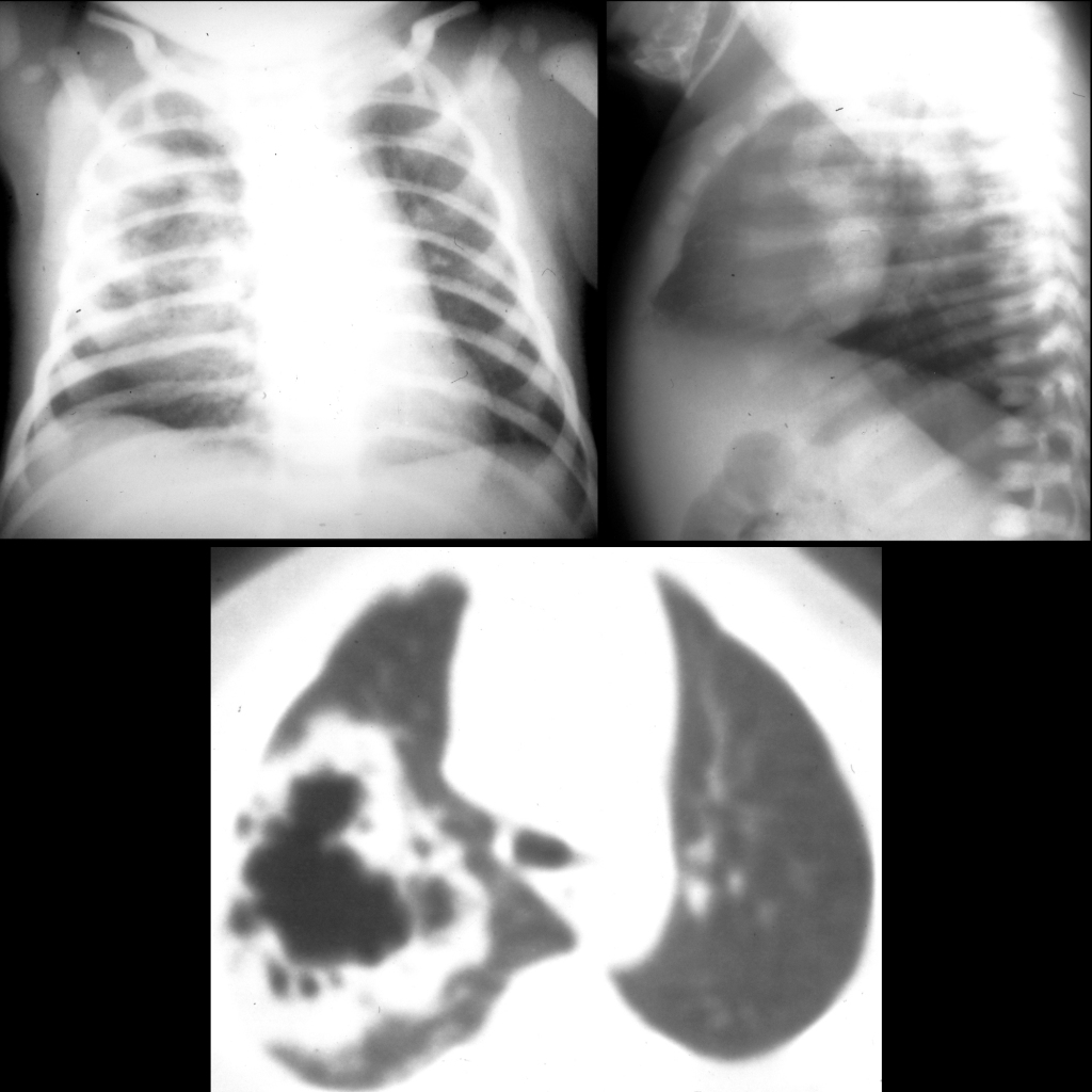
Premature newborn now 2 months old with respiratory distress

CXR and CT of pneumatocele
CXR (above) and axial CT without contrast of the chest (below) shows diffuse interstitial thickening and a large, round, unilocular thin-walled cyst in the posterobasal aspect of the right lower lobe. The bronchus of the posterobasal segment of the right lower lobe was seen to extend into it.

The diagnosis was pneumatocele due to positive pressure ventilation in a patient with chronic lung disease.
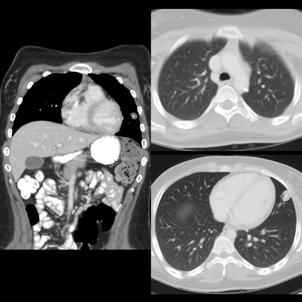
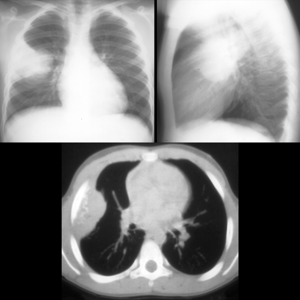
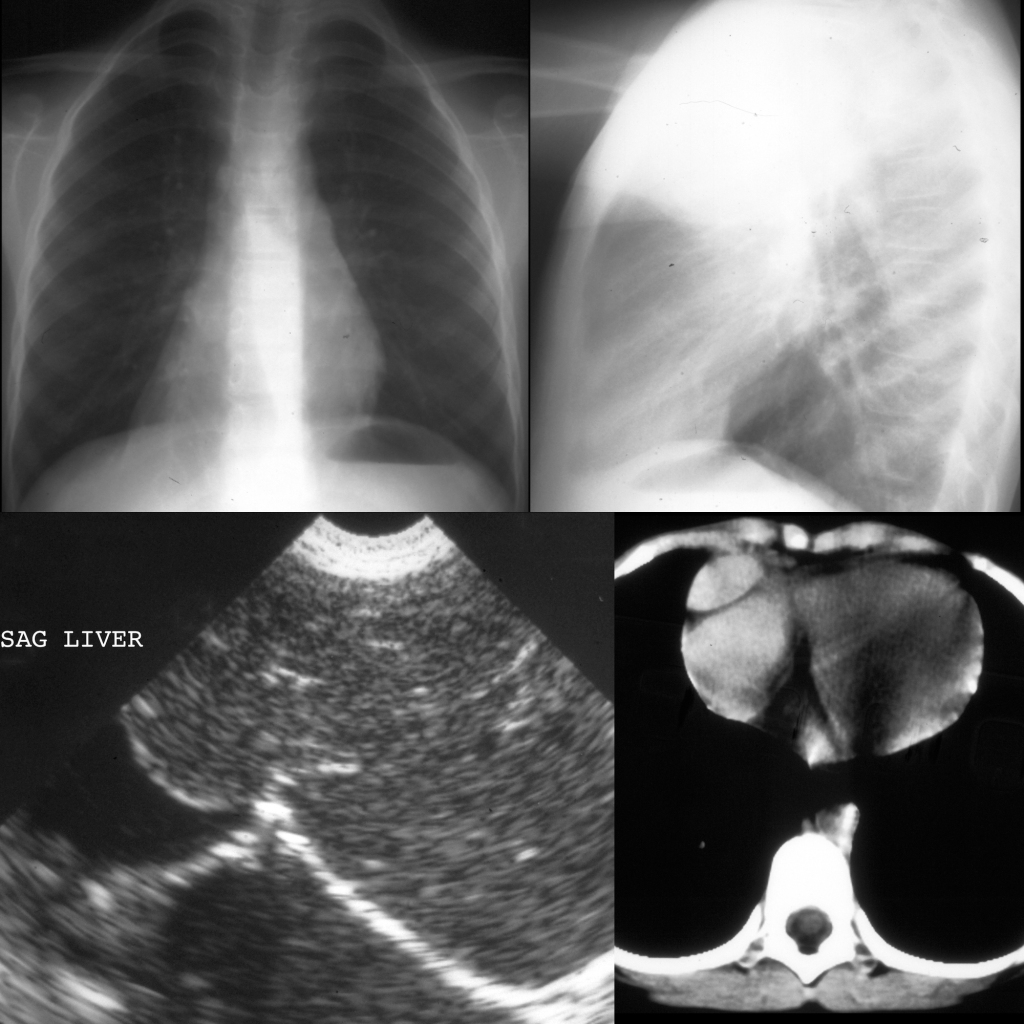
School ager with substernal distress associated with exercise

CXR and US and CT of Morgani hernia
CXR AP and lateral (above) shows an anteromedial soft tissue density in the right cardiophrenic angle obscuring the right heart border. Sagittal US of the chest performed anteriorly and just to the right of the midline (below left) shows liver extending above the level of the diaphragm. Axial CT with contrast of the chest (below right) shows liver in the right cardiophrenic angle just to the right of the heart in the right hemithorax.

The diagnosis was a right-sided Morgagni hernia containing liver.
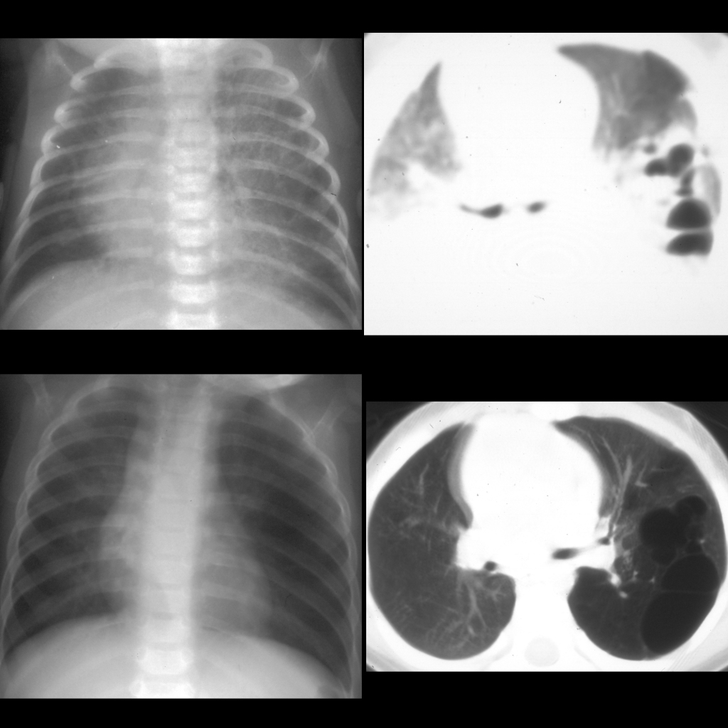
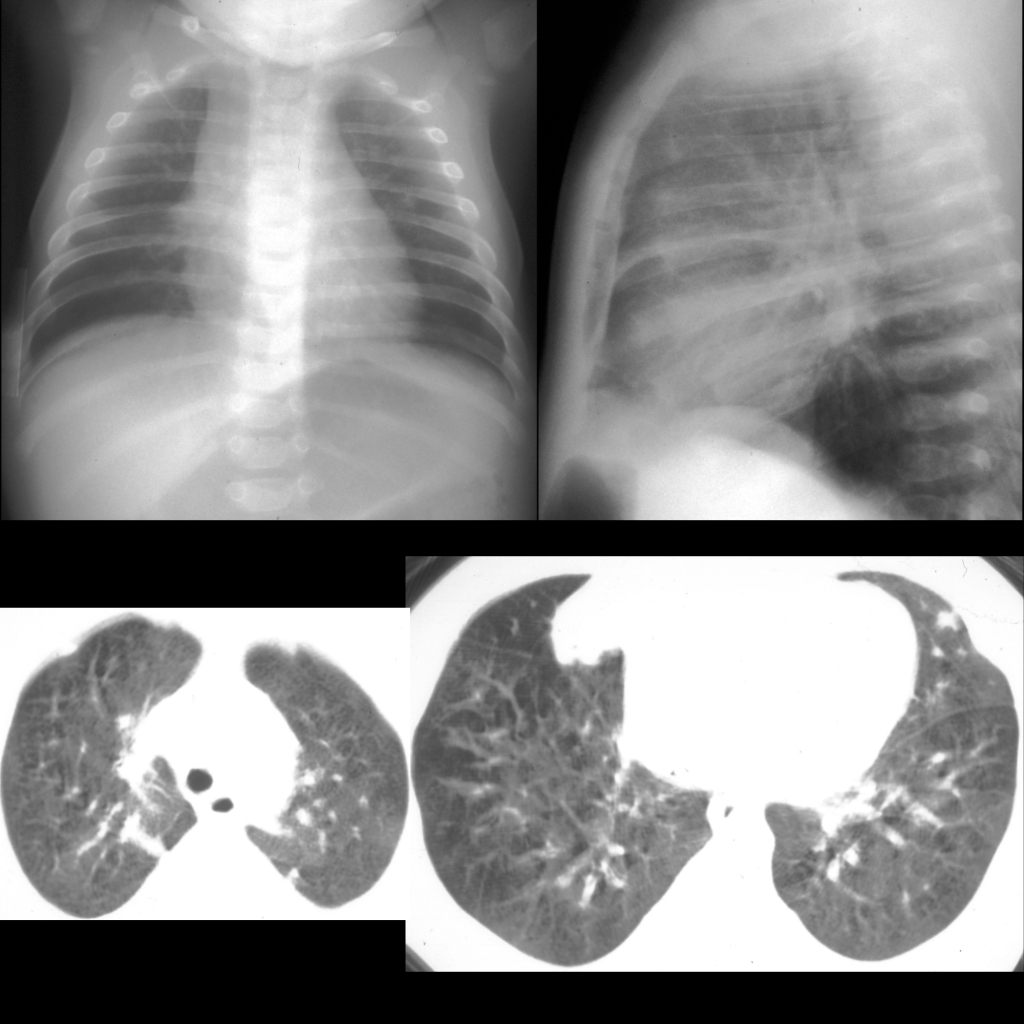
Newborn with an echogenic left lower lung mass on prenatal ultrasound

CT of congenital pulmonary airway malformation Type I
CXR at birth (above left) shows an ill-defined large mass in the lower left chest obscuring the heart border and causing mediastinal shift to the right. Axial CT without contrast of the chest at birth (above right) shows the left chest mass to contain multiple cysts that are filled with air and fluid. CXR at 10 months (below left) shows the large lower left chest mass to now be hyperlucent. Axial CT without contrast of the chest at 10 months (below right) shows the left chest mass to contain multiple large air-filled cysts.

The diagnosis was congenital pulmonary airway malformation Type I.
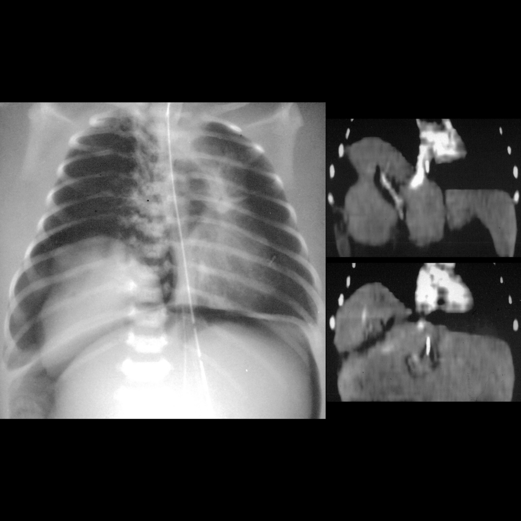
Newborn with respiratory distress and new abdominal distension

CT of congenital diaphragmatic hernia
CXR (left) shows massive pneumoperitoneum and right pneumothorax which outlines a mass in the lower right chest which is causing mediastinal shift to the left. Coronal CT with contrast of the chest (above right) shows liver herniating through a lateral defect in the diaphragm into the lower right chest and lying above the right diaphragm (below right).

The diagnosis was congenital diaphragmatic hernia containing liver as outlined by pneumothorax and pneumoperitoneum.
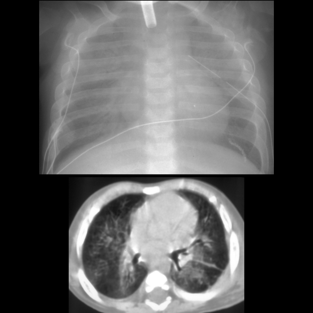
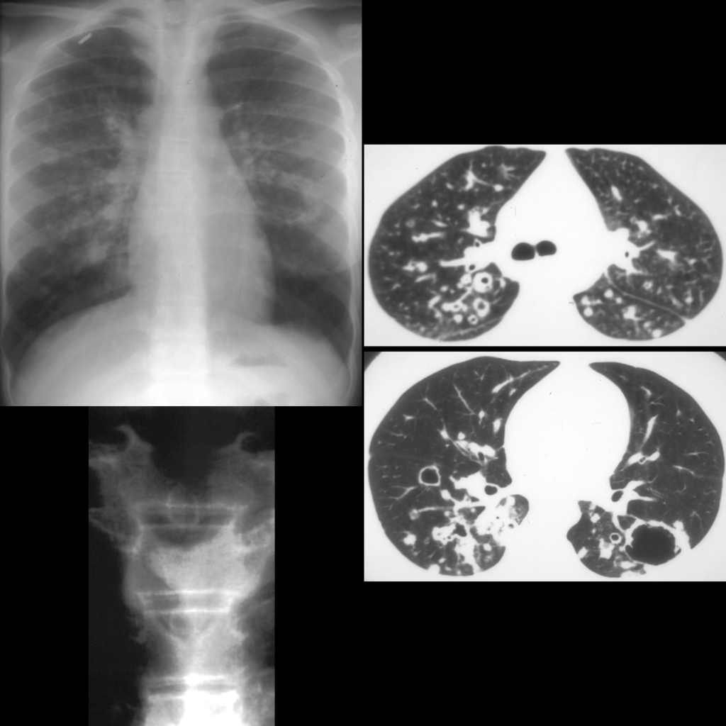
Teenager on chemotherapy with fever

CXR and CT of invasive aspergillosis
CXR on day 8 of the fever (above left) shows opacities in the left upper lobe and right middle lobe. Axial CT without contrast of the chest on day 13 of the fever (above right) shows parenchymal opacities surrounded by irregular ground glass density (halo sign). CXR on day 20 of the fever (below left) shows interval cavitation of the bilateral lung lesions which on axial CT with contrast of the chest on day 34 of the fever (below right) shows central necrosis in the lesions which is detached from the wall (air crescent sign).

The diagnosis was invasive aspergillosis.